A system for histological examination of tissue specimens

a tissue specimen and histological examination technology, applied in the field of histological examination systems of tissue specimens, can solve the problems of ineffective auditing methods of users, inability to effectively consult with colleagues about specimens, and inefficient operation of microscopes, so as to increase the visibility of projected image light

Active Publication Date: 2020-07-02
AUGMENTIQS MEDICAL LTD
View PDF5 Cites 8 Cited by
  • Summary
  • Abstract
  • Description
  • Claims
  • Application Information

AI Technical Summary

Benefits of technology

The patent text describes an apparatus that improves the visibility of images generated by a digital image projector when viewed through a microscope. The apparatus includes a shutter that blocks at least some of the light from the objective of the microscope, making the image darker and easier to see. This helps to improve the overall quality of the image and make it easier to analyze and investigate.

Problems solved by technology

However, for a busy microscope user, such as, but not limited to a pathologist, the operation of a microscope is not necessarily efficient in terms of reaching the best diagnosis, or completing a case report.
Moreover, there is currently no effective method for auditing work performed by the user, nor is the user able to effectively consult with colleagues about the specimen.
While today, many microscopes enable imaging cameras within the third ocular that are used to capture images into a personal computer station where they can be further analyzed and saved as reports, it has been found that many microscope users do not generally use these imaging cameras due to their slowing down the workflow.
This is a time-consuming situation which does not lend well to improving specimen diagnosis workflow.
Moreover, lack of routine photography in practical clinical settings limits generation of clinical data arranged in databases that is a critical component of development of automated diagnostics and / or diagnostics support algorithms.
Within the limitations of the present technology the scanning / information retrieval takes substantially longer time as compared to manual microscopy.
This drawback imposes significant hurdles for practical deployment of those systems.

Method used


Image

  • A system for histological examination of tissue specimens
  • A system for histological examination of tissue specimens
  • A system for histological examination of tissue specimens

Examples


Embodiment Construction

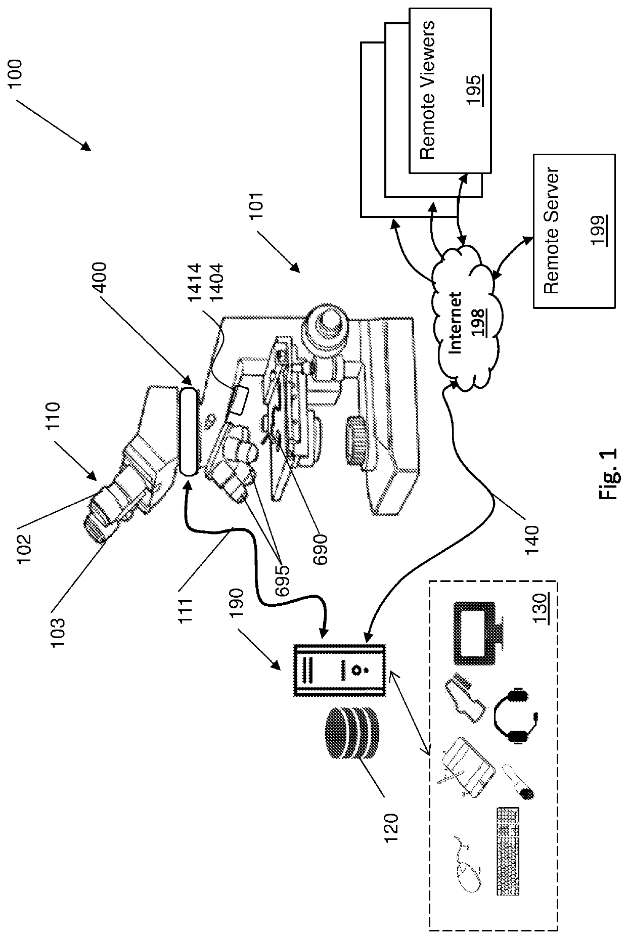
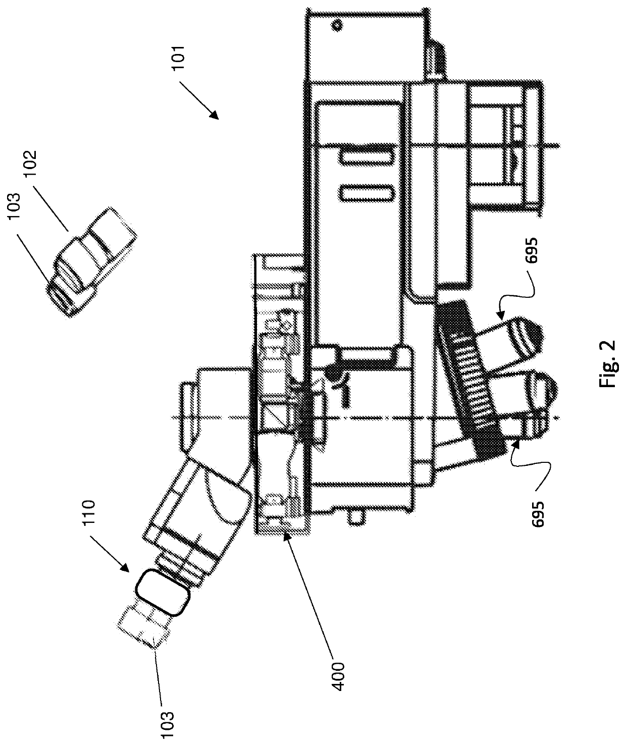
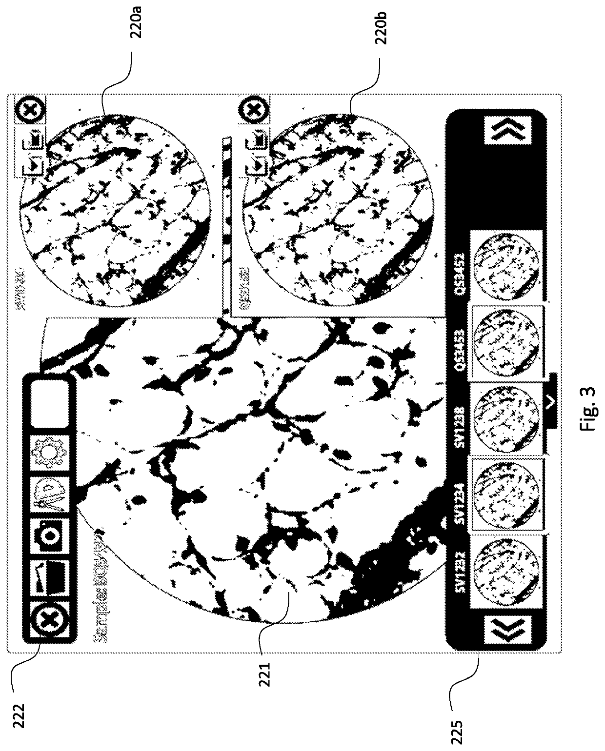
[0023]According to another aspect of the presently disclosed subject matter, there is provided a system retrofitting an optical microscope by inserting an electro-optical unit into the optical accessory bay of the microscope or other suitable port within the image optical path of the microscope, such that the system enables viewing of the optical imaging plane, acquisition of digital images from the optical imaging plane, and a display of pointers and digital information as an overlay on the optical image of the specimen.

[0024]In an exemplary embodiment, the electro-optical unit is inserted at or near the Fourier plane of the optical train. The electro-optical unit comprises an image capture and image projection units.

[0025]In an exemplary embodiment, the electro-optical unit has a form-factor that allows it to be inserted into the optical train of a standard microscope, for example in the filter's bay.

[0026]In another exemplary embodiment, the standard camera port of the microscope...


PUM


Abstract

An interactive apparatus, system and method for image capturing and projecting is integrated into an optical microscope. An image capturing and projecting unit is operatively connected to a processing unit, the processing unit configured to: (a) receive user generated data; and (b) overlay the user generated data onto an optically viewed image visible through the eyepiece.

Description

FIELD OF THE INVENTION[0001]The present invention relates to the field of manual and automated microscopy, and particularly to a microscope having an eyepiece, an illumination beam path serving to illuminate a specimen, and a detection beam path that images the specimen, where additional data can be overlaid within the eyepiece, and methods for operations thereof.BACKGROUND OF THE INVENTION[0002]Microscopes have been in use for over 200 years, with few conceptual changes to their design and use over this period. However, for a busy microscope user, such as, but not limited to a pathologist, the operation of a microscope is not necessarily efficient in terms of reaching the best diagnosis, or completing a case report.[0003]Currently when beginning a specimen examination, a microscope user looks through the eyepieces in order to view the specimen. The user then makes a decision or diagnosis based on characteristics or features of the specimen according to their best judgement. In othe...

Claims


Application Information

Patent Timeline
IPC IPC(8): G06T11/00G02B21/18H04N5/225G06F3/01
CPCG02B21/18H04N5/2254H04N5/2256G06F3/013G06T11/00G02B21/361G02B21/362G02B21/368G16H10/40H04N23/55H04N23/56
Inventor SIEGEL, GABRIELREGELMAN, DAN VADIM
Owner AUGMENTIQS MEDICAL LTD
Why Eureka
Try Eureka