Uncaging stent
a stent and stent technology, applied in the field of uncaging stents, to achieve the effects of high radial strength, high stent strength, and loss of scaffold structural integrity
- Summary
- Abstract
- Description
- Claims
- Application Information
AI Technical Summary
Benefits of technology
Problems solved by technology
Method used
Image
Examples
example 1
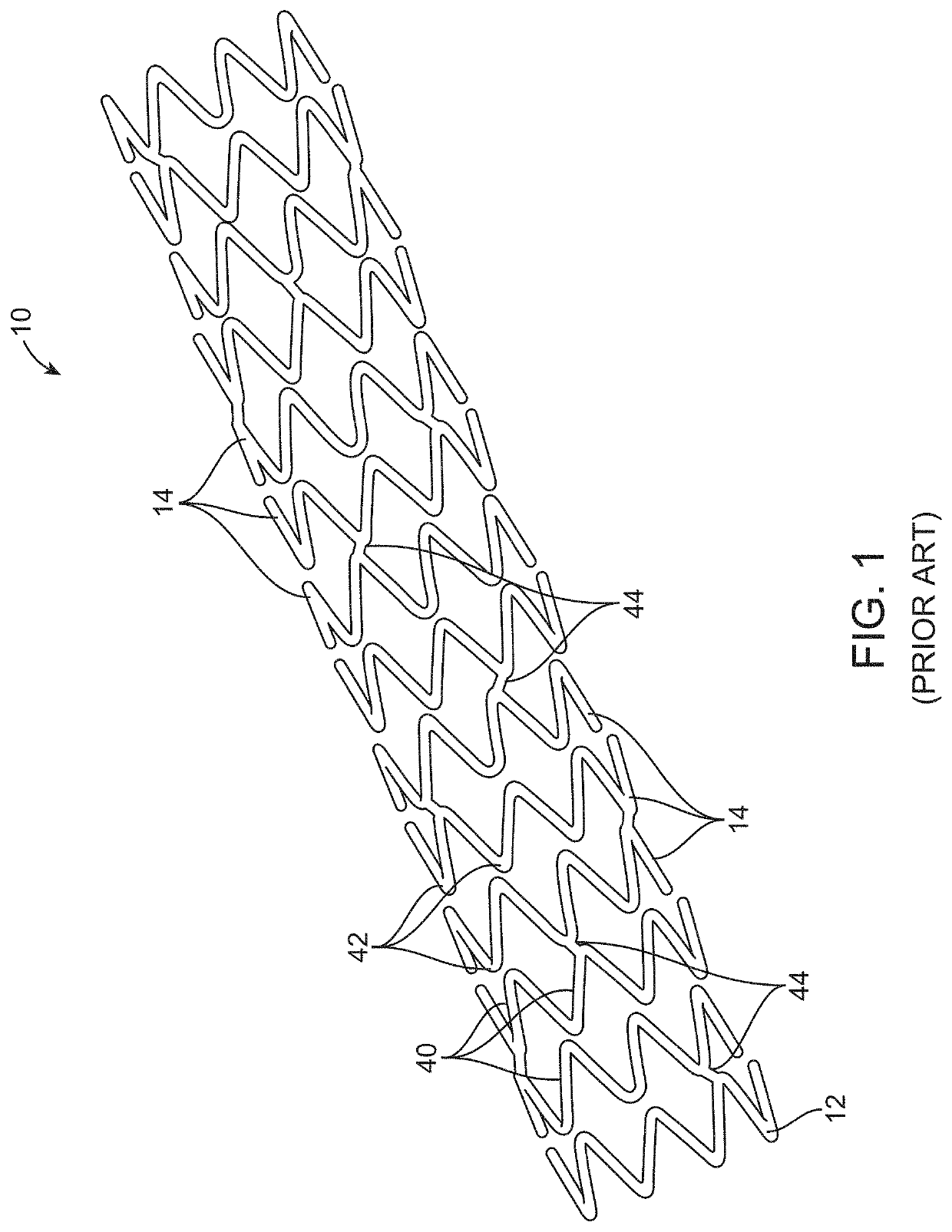
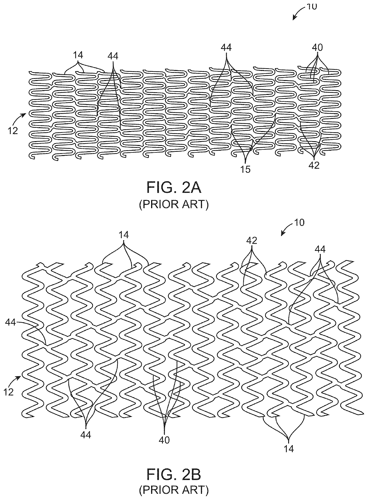
[0886]Vasomotion evaluation by IVUS: A porcine animal having a control non-degradable plastically deformable stent (DESyne, Elixir Medical Inc. n=2) and a test stent of (PR153RG, n=4) having a 6-crown 3-link non-degradable plastically deformable stent patterned to have three evenly spaced separation regions per ring, with the S shaped axial links connecting adjacent rings was tested and followed up at the 3 month time point. The test devices were coated with a fast degrading (from 1-3 months) lactide copolymer covering the separation regions (including the inner, sides, and outer surfaces of the separation regions and the space within the separation regions) and covering the stent surfaces (luminal, abluminal and two side surface). The stents were further coated with a top coat of Novolimus and a lactide copolymer drug matrix. The abluminal and luminal coating thickness was about 10 microns. The test stents and the control stents (DESyne, Elixir Medical Inc.) were implanted in the c...
example 2
[0889]Finite Element Analysis Model testing for the different Tests listed below were conducted on the stent of the present invention. The mock artery had a wall thickness of 0.010″, regardless of diameter, and an inner diameter match the outer diameter of the stent. The artery wall material was modeled as a linear elastic material with a Poisson's ratio of 0.45, and exhibits a 5% diametric compliance when pressurized with 100 mmHg on the inner surface of the artery. The Young's modulus varied between 300 and 700 PSI, depending on the arterial diameter. The Stent material was a linear elastic material having a Young's modulus of between 36,000,000 and 39,000,000 PSI (36e6 to 39e6 PSI) and a Poisson's ratio of 0.3. FEA model used FEMAP and Nastran software and consisted of arrangements of 10 node modified Tet elements at a density shown to give results accurate to less than + / −5%. The minimum mesh density used in this analysis had 1 layer of elements through the thickness of the arte...
example 3
[0890]Three point Bend Test FIG. 15: A stent was patterned from Stent Material having a thickness of 75 microns, having a serpentine ring with each ring having 3 separation regions. The stent was in an expanded configuration of diameter 3.5 mm and is connected to a mock artery. Fixtures were connected at each end and in the middle. The minimum separation between the end connectors was 11 mm. The fixture at one end of the model was allowed to pivot along a line as shown below, but was otherwise fixed against movement. The fixture at the opposite end could pivot and move relative to the first end in the direction of the axis of the stent. The fixture on top in the middle was then displaced by 1 mm toward the stent, bending the stent and mock artery. Linear elastic analysis was used, resulting in an estimated Force of 1.4N prior to the formation of discontinuities, which dropped to 0.4N after formation of discontinuities. The stent of the current invention with discontinuities in the s...
PUM
| Property | Measurement | Unit |
|---|---|---|
| curvature | aaaaa | aaaaa |
| reversing curvature | aaaaa | aaaaa |
| direction of curvature | aaaaa | aaaaa |
Abstract
Description
Claims
Application Information
 - R&D
- Intellectual Property
- Life Sciences
- Materials
- Tech Scout
- Unparalleled Data Quality
- Higher Quality Content
- 60% Fewer Hallucinations
Browse by: Latest US Patents, China's latest patents, Technical Efficacy Thesaurus, Application Domain, Technology Topic, Popular Technical Reports.
© 2025 PatSnap. All rights reserved.Legal|Privacy policy|Modern Slavery Act Transparency Statement|Sitemap|About US| Contact US: help@patsnap.com


