
Method for the Analysis of Glycosaminoglycans, and Their Derivatives and Salts by Nuclear Magnetic Resonance

a glycosaminoglycan and nuclear magnetic resonance technology, applied in the direction of analysis using nuclear magnetic resonance, measurement using nmr, instruments, etc., can solve the problems of inability to obtain structural information directly from this area, the complete characterization of heparins and low molecular weight heparins is currently a challenge, and the difficulty of obtaining structural information prior to this area

Active Publication Date: 2020-08-27
LAB FARM ROVI SA
View PDF0 Cites 1 Cited by
  • Summary
  • Abstract
  • Description
  • Claims
  • Application Information

AI Technical Summary

Benefits of technology

This approach provides high specificity, accuracy, and repeatability in quantifying saccharide residues, enabling the identification of GAGs and their manufacturing processes, thus addressing the limitations of prior art by facilitating reliable quality control and biosimilarity assessments.

Problems solved by technology

Currently, no prior art NMR method nor corresponding composition for the highly accurate quantification of individual saccharides in GAG's and heparin molecules exist.
However, given the high degree of microheterogeneity and polydispersity of these compounds, the complete characterization of heparins and low molecular weight heparins is currently still a challenge.
The region between 2.8-4.5 ppm comprises the majority of the saccharide ring signals and has a high degree of overlapping between them, which makes it difficult to extract structural information directly from this area.
However, the absence of precise methods that consider and control both the experimental methods and the processing and evaluation of the spectra means that measurements made on identical samples in various laboratories may significantly differ (“Validation of quantitative NMR”, Malz F. and Jancke H. in Journal of Pharmaceutical and Biomedical Analysis (2005), 38, 813-823).
The complexity of the nuclear magnetic resonance spectra of glycosaminoglycans in general, and heparins and low molecular weight heparins and their derivatives in particular, has meant that to date no specific validation methods have been developed which allow quantification of its characteristic signals, and therefore, the suitable characterization and differentiation of these compounds.
In other words, prior art NMR methods have not successfully achieved highly accurate quantitation of each of the individual saccharide residues present GAG's and heparins.
The absence of these methods prevents the suitable comparability between identical samples studied and assessed under not suitably established experimental conditions.

Method used


Image

  • Method for the Analysis of Glycosaminoglycans, and Their Derivatives and Salts by Nuclear Magnetic Resonance
  • Method for the Analysis of Glycosaminoglycans, and Their Derivatives and Salts by Nuclear Magnetic Resonance
  • Method for the Analysis of Glycosaminoglycans, and Their Derivatives and Salts by Nuclear Magnetic Resonance

Examples


example 1

1H NMR of Enoxaparin Sodium

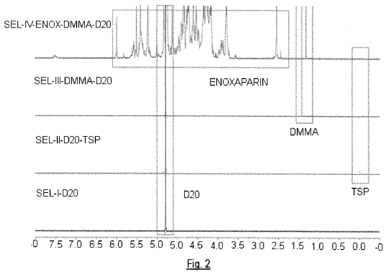
[0219]Enoxaparin sodium (50 mg) are dissolved in 500 μL of a D2O-TSP (solution B) solution. Then 100 μL of DMMA solution (solution C) are added. The resulting solution is introduced in a 5 mm diameter tube.

[0220]The resulting DMMA concentration in the solution is 1.5 mM. The experiments are performed on a Bruker AVIII-800 nuclear magnetic resonance spectrometer. The main signals identified are as follows:

SignalChemical shift, ppmH4 ΔU2S5.992H4 ΔU25.825H1 1,6-AnA5.616H1 ANS(-G)5.585H1 1,6-AnM5.569H1 ΔU2S5.509H1 ANS6S5.405H1 I2S5.228H1 I5.012H5 I2S4.836H1 G4.628H6 ANS6S4.344H6′ ANS6S4.210H3 ANS3.670H2 ANS3S3.395H2 ANS3.293NAc2.047DMMA1.320TSP0.069

[0221]Once the values of the integrals of the signals both of DMMA and the rest of the residues have been obtained, the normalized values are obtained of said residues dividing the value of its integrals by the value of the integral corresponding to the DMMA signal. This normalization can be performed, because the c...

example 2

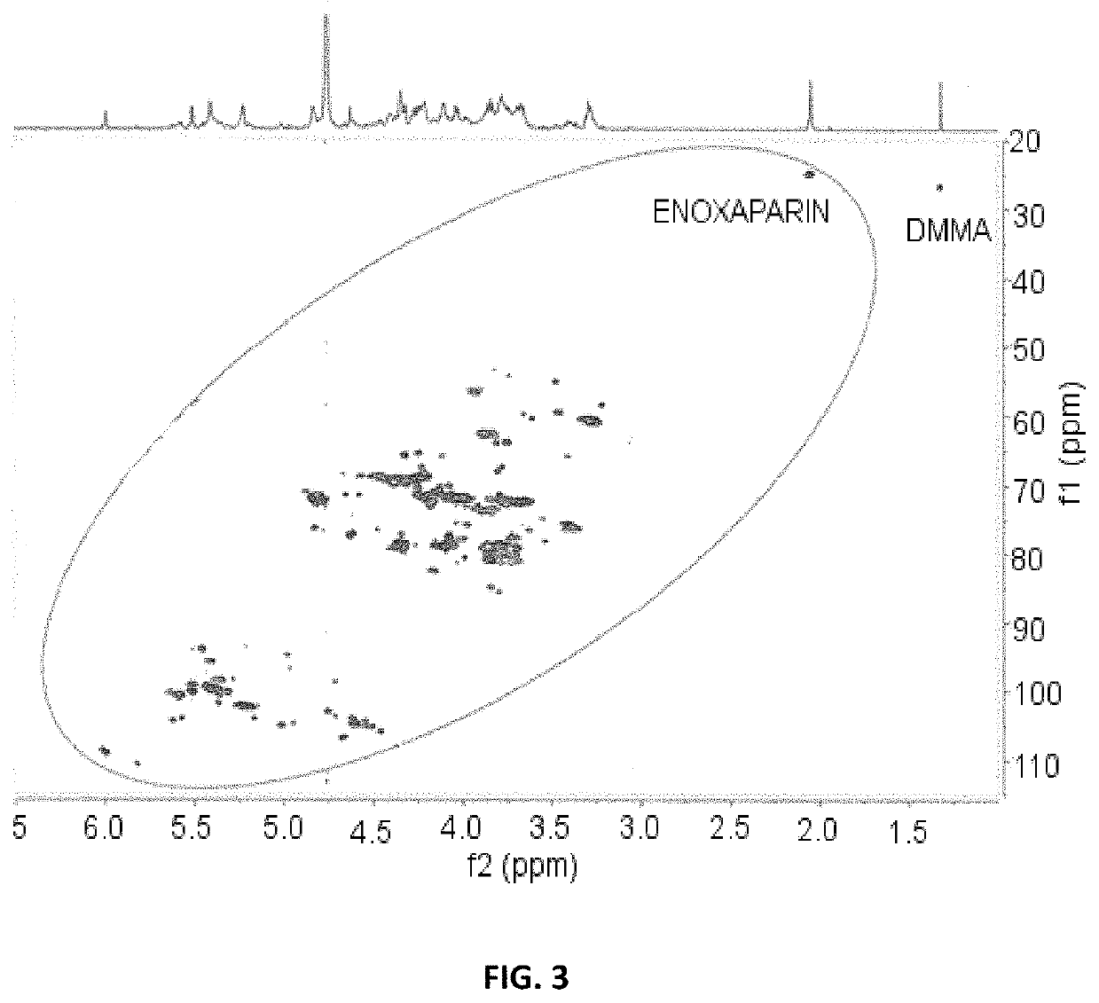
[0228]The same solution used in Example 1 is used to perform the study by 1H-13C HSQC. The main signals identified are as follows:

Signalδ13C, ppmδ1H, ppmSignalδ13C, ppmδ1H, ppmC4—H4 ΔU110.715.82C3—H3 Gal85.453.78C4—H4 ΔU2S108.975.99C3—H3 Gal84.853.83C1—H1 G / / Gal106.624.66C4—H4 ANS6S(-G) / / 80.943.84C1—H1 Xyl105.794.45ANS6S redC1—H1 G(-ANAc)105.134.50C2—H2 I2S78.534.34C1—H1 I(-A6S)104.945.01C3—H3 Xyl77.823.72C1—H1 G(-ANS)104.774.60C2—H2 ΔU2S77.424.62C1—H1 I(-A6OH)104.674.94C2—H2 G(-AN6S)75.703.40C1—H1 Gal104.304.54C3—H3 ANS6S (-G)72.473.66C1—H1 1,6-an.A104.225.61C3—H3 ANS6S red72.293.77C1—H1 G(-ANS3S)103.914.61C5—H5 I2S72.014.83C1—H1 1,6-an.M103.915.57C3—H3 I2S71.874.21C1—H1 ΔU103.885.16C5—H5 ANS6S(-G)71.724.09C1—H1 G2S102.994.75C5—H5 MNS6S red70.984.15C1—H1_I2S102.095.22C5—H5 ANS6S red70.644.12C1—H1 I2S(-1,6-101.595.36C6—H6 1,6-an.A / / 67.533.77an.M)1,6-an.MC1—H1 ANS(-G)100.505.58C5—H5 Xyl65.894.12C1—H1 ANAc100.235.31C5—H5 Xyl65.863.40C1—H1 ΔU2S100.185.51C3—H3 ΔU2S65.754.32C1—H1 ANS...

example 3

Study by 1H NMR of Bemiparin Sodium

[0232]The main 1H chemical shift signals identified were as follows:

SignalChemical shift, ppmH4 ΔU2S5.992H4 ΔU5.825H1 1,6-AnA5.616H1 ANS(-G)5.585H1 1,6-AnM5.569H1 ΔU2S5.509H1 ANS6S5.405H1 I2S5.228H1 I5.012H5 I2S4.836H1 G4.628H6 ANS6S4.344H6′ ANS6S4.210H3 ANS3.670H2 ANS3S3.395H2 ANS3.293NAc2.047DMMA1.320TSP0.069

[0233]The quantification of the characteristic and well-differentiated signals of bemiparin sodium (generally those corresponding to the anomeric or target protons, H1) are shown in the following table, with the values of relative proportions observed for a series of six samples.

SignalChemical shift, ppmRelative proportion, %H4 ΔU2S5.993.7-5.7H4 ΔU5.820.2-2.5H1 1,6-an.A5.620.5-2.5H1 1,6-an.M5.572.5-6.0H1 ΔU2S5.51 7.0-10.7H1 ANS6S5.4019.0-21.3H1 I2S5.2313.8-18.5H2 ANS3.2918.7-26.3NAc2.05 9.4-14.4


PUM


Abstract

A nuclear magnetic resonance method of analysis for glycosaminoglycans in general, and of heparins and low molecular weight heparins and their derivatives in particular, is provided. The method allows for their identification and for relative quantification of respective characteristic signals by 1H-NMR and / or 1H-13C HSQC.

Description

CROSS-REFERENCE TO EARLIER FILED APPLICATIONS[0001]The present application claims the benefit of and is a continuation of application 16 / 251909 filed Jan. 18, 2019, which is a continuation-in-part of international application PCT / EP2017 / 068285 filed Jul. 19, 2017, which claims the benefit of European application EP 16382350.3 filed Jul. 19, 2016, the entire disclosures of which are hereby incorporated by reference.FIELD OF THE INVENTION[0002]The present invention concerns an analytical method employing nuclear magnetic resonance (1H-NMR, 13C-NMR, and / or 1H-13C-HSQC) for the characterization of glycosaminoglycans (GAG's), derivatives thereof, and / or salts thereof. The method allows for qualitative and quantitative analysis of the saccharides comprising said GAG's.BACKGROUND OF THE INVENTION[0003]Nuclear magnetic resonance (NMR) spectroscopy is one of the most important and widespread analytical techniques used in the characterization of glycosaminoglycans in general, and heparins and...

Claims


Application Information

Patent Timeline
Patent Type & Authority Applications(United States)
IPC IPC(8): G01N24/08G01R33/46G01N33/50
CPCG01N24/087G01R33/4625G01N24/08G01N33/50G01R33/4633
Inventor FRANCO RODRIGUEZ, GUILLERMOGUTIERRO ADURIZ, IBON
Owner LAB FARM ROVI SA
Learn More