
DNA targets as tissue-specific methylation markers

Pending Publication Date: 2020-10-29
YISSUM RES DEV CO OF THE HEBREWUNIVERSITY OF JERUSALEM LTD +1
View PDF0 Cites 0 Cited by
  • Summary
  • Abstract
  • Description
  • Claims
  • Application Information

AI Technical Summary

Benefits of technology

The present invention provides a method for identifying the methylation status of double-stranded, cell-free DNA molecules in a specimen, which can be used to detect the presence or absence of a specific cell type or tissue in the specimen. This method involves analyzing the methylation status of at least two methylation sites on a continuous nucleotide sequence of the DNA molecule. The invention also provides a kit for identifying the source of DNA in a sample and a method for classifying diseases associated with tissue damage, such as sepsis, lupus, and HIV. The DNA molecule analyzed can be no longer than 300 base pairs and the methylation sites can be not more than 300 bp apart on a single strand.

Problems solved by technology

However in these cases, the source of elevated cfDNA is unknown, greatly compromising the utility of cfDNA as a diagnostic or prognostic tool.
Note that since the approach relies on normal, stable markers of cell identity, it cannot identify the nature of the pathology (e.g. distinguishing cfDNA derived from dead tumor cells or dead wild type cells due to trauma or inflammation in the same tissue).

Method used


Image

  • DNA targets as tissue-specific methylation markers
  • DNA targets as tissue-specific methylation markers
  • DNA targets as tissue-specific methylation markers

Examples


example 1

Analyzing Methylation Patterns of Cardiac Markers

[0291]Materials and Methods

[0292]Clinical Samples:

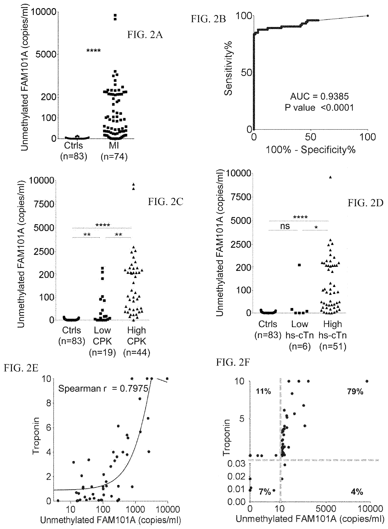
[0293]Cardiac biomarkers used were troponin T and CPK.

[0294]Identification of Cardiac Methylation Markers:

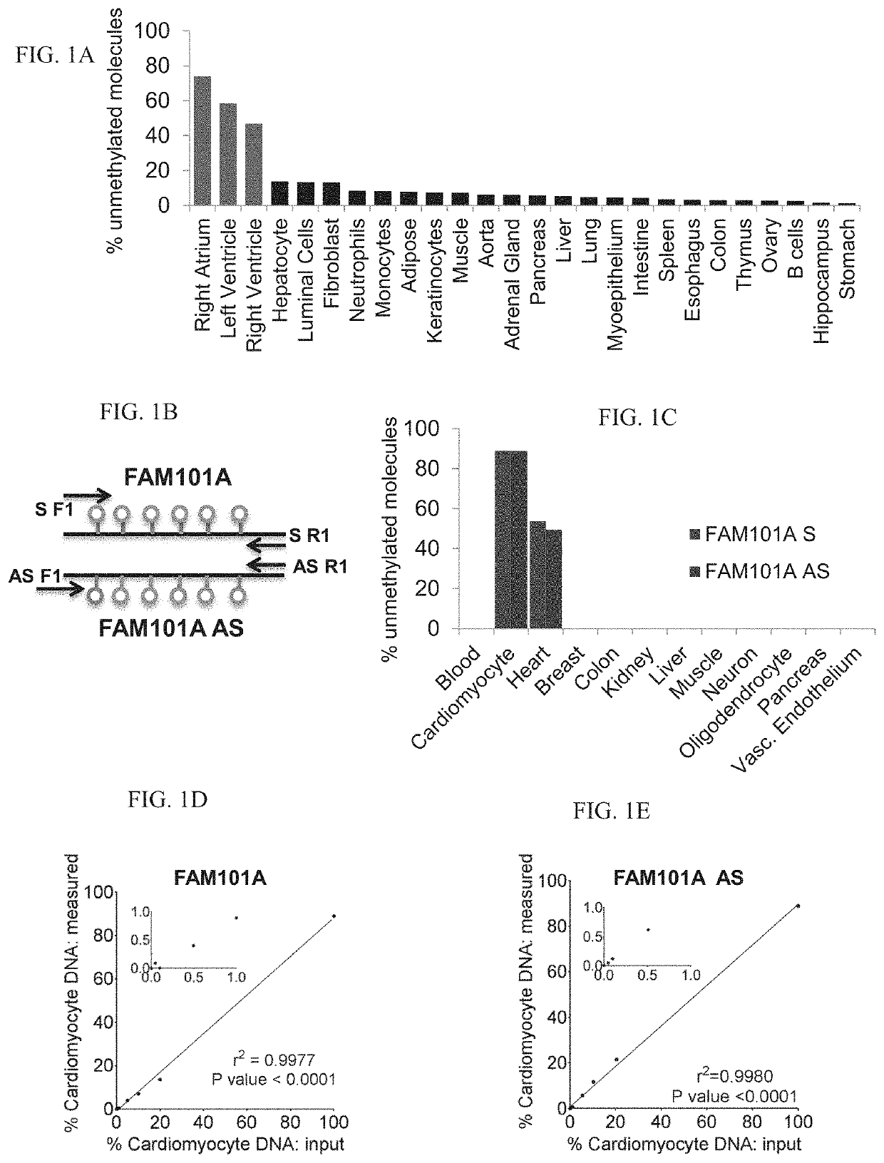
[0295]Tissue-specific DNA methylation markers were selected after a comparison of publically available DNA methylation datasets generated by whole-genome bisulfite sequencing (Roadmap Epigenomics). The fragment of FAM101A used as a cariomyocyte-specific marker is located in chromosome 12, coordinates124692462-124692551.

[0296]Cfdna Analysis:

[0297]Blood samples were collected in EDTA tubes, and centrifuged within 2 hours to separate plasma from peripheral blood cells: first at 1500 g for 10 min, and then at 3000 g for 10 min to remove any remaining cells. Plasma was then stored at −80° C.

[0298]cfDNA was extracted using the QIAsymphony SP instrument and its dedicated QIAsymphony Circulating DNA Kit (Qiagen) according to the manufacturer's instructions. DNA concentration was measured u...

example 2

Analyzing Hepatic Cell Methylation Signatures

[0327]The clinical standard to assess liver damage is serum measurement of alanine aminotransferase (ALT) and aspartate aminotransferase (AST), reflecting hepatocyte injury leading to release of these enzymes into the blood. While in extensive clinical use, these tests do have important limitations. First, the enzymes are not absolutely hepatocyte-specific. AST is expressed in cardiac and skeletal muscle, kidney, brain, pancreas, lung, leukocytes and erythrocytes, while ALT is primarily present in the liver and kidneys, with low amounts in the heart and skeletal muscle. Second, liver enzymes do not always reflect the full burden of disease and in various hepatic pathologies have been shown to be insufficient. A possible reason for this is that the enzymes could be released from dying as well as reversibly injured cells, and therefore do not indicate the exact nature of damage to the liver, nor the number of injured cells. Third, ALT and A...

example 3

List of Additional Identified Targets

[0368]A list of identified targets is provided in Table 1 and 2 herein below. The targets can be used to identify a cell type of the listed organ. It will be appreciated that the sequences provided are 500 base pairs. Preferably the target sequence which is amplified comprises the nucleotides CG which are at position 250 and 251 of each of these sequences and additional nucleotides up and / or down-stream of this site.

TABLE 1SEQ IDOrganNameNO:AcinarCPA12AcinarLMF23AcinarNCLN4AcinarBRF15AcinarFRY6AstrocytesHDAC47AstrocytesAGAP18AstrocytesASTI9AstrocytesPRDM10AstrocytesFOXP411AstrocytesKIAA12AstrocytesPRDM213AstrocytesWWOX14B cellsLRP515B cellsSORL116B cellsTRPV117BETAINSh18BETAMTG119BETAZC3H320BETALeng821BETAFbxw822BETAFbxl1923BloodLoc1 / AGAP224BloodPTPRCAP25BRAINMAD1L126BRAINPTPRN227BRAINWM128BRAINMBP29BRAINNUMBLE30BRAINLRRN331BRAINcg097832BRAINZNF23833BrainWB134BrainUBE4B35BreastKRT1936BreastLMX1B37BreastZNF29638CD8 cellsCD8A39CD8 cellsCD8A anti40C...


PUM

PropertyMeasurementUnit
Massaaaaaaaaaa
Massaaaaaaaaaa
Massaaaaaaaaaa

Abstract

A method of ascertaining the methylation status of a double-stranded, cell-free DNA molecule in a specimen is disclosed. The method comprises ascertaining the methylation status of at least two methylation sites of the same double-stranded cell-free DNA molecule, wherein said double-stranded, cell-free DNA molecule comprises a nucleotide sequence which comprises no more than 300 base pairs and is comprised in a sequence as set forth in any one of SEQ ID NOs: 2-117 or 121-177.

Description

FIELD AND BACKGROUND OF THE INVENTION[0001]The present invention contemplates novel target sequences that can be used as tissue-specific methylation markers.[0002]It has been known for decades that plasma contains small fragments of cell-free circulating DNA (cfDNA) derived from dead cells (on average 1000 genome equivalents per ml). While the mechanisms underlying the release and clearance of cfDNA remain obscure, the phenomenon is rapidly being exploited for a variety of applications with clinical relevance. The recognition that fragments of fetal DNA travel briefly in maternal circulation has opened the way for next generation sequencing (NGS)-based prenatal testing to identify fetal trisomies and other genetic aberrations, potentially replacing amniocentesis. In cancer biology, tumors are known to release DNA (including tumor-specific somatic mutations) into the circulation, providing means for liquid biopsies to monitor tumor dynamics and genomic evolution. In addition, cfDNA h...

Claims


Application Information

Patent Timeline
IPC IPC(8): C12Q1/6883C12Q1/686
CPCC12Q2600/154C12Q2600/112C12Q1/6883C12Q1/686C12Q2600/118C12Q1/6827C12Q2523/125C12Q2531/113C12Q2563/159
Inventor DOR, YUVALSHEMER, RUTHGLASER, BENJAMINMAGENHEIM, JUDITHNEIMAN, DANIELWERMAN, RONIZEMMOUR, HAIMOSS, JOSHUAFOX, ILANAPIYANZIN, SHEINA
Owner YISSUM RES DEV CO OF THE HEBREWUNIVERSITY OF JERUSALEM LTD
Learn More