Compositions and methods to increase muscular strength
a technology applied in the field of compositions and methods for increasing muscle strength, can solve the problems of limiting the amount of force people can exert, affecting their day-to-day mobility and basic functions, and people have limited mobility, so as to improve external force and increase muscle for
- Summary
- Abstract
- Description
- Claims
- Application Information
AI Technical Summary
Benefits of technology
Problems solved by technology
Method used
Image
Examples
example 1
iated Skeletal Muscle Tissue and Contractility
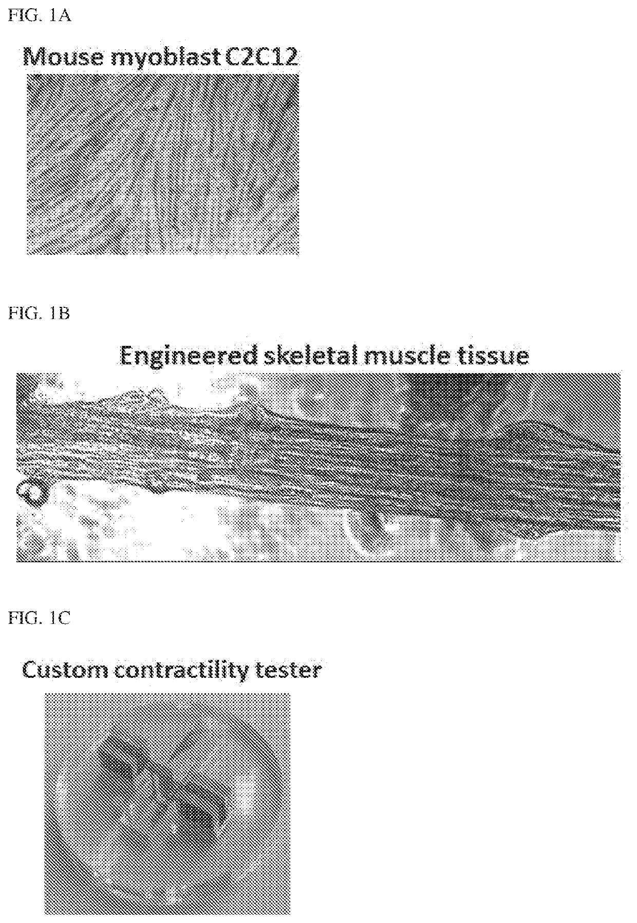
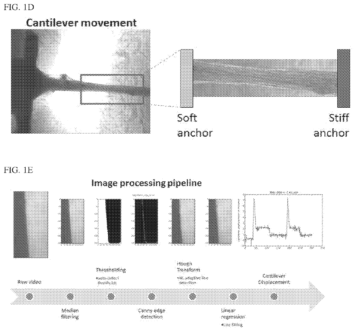
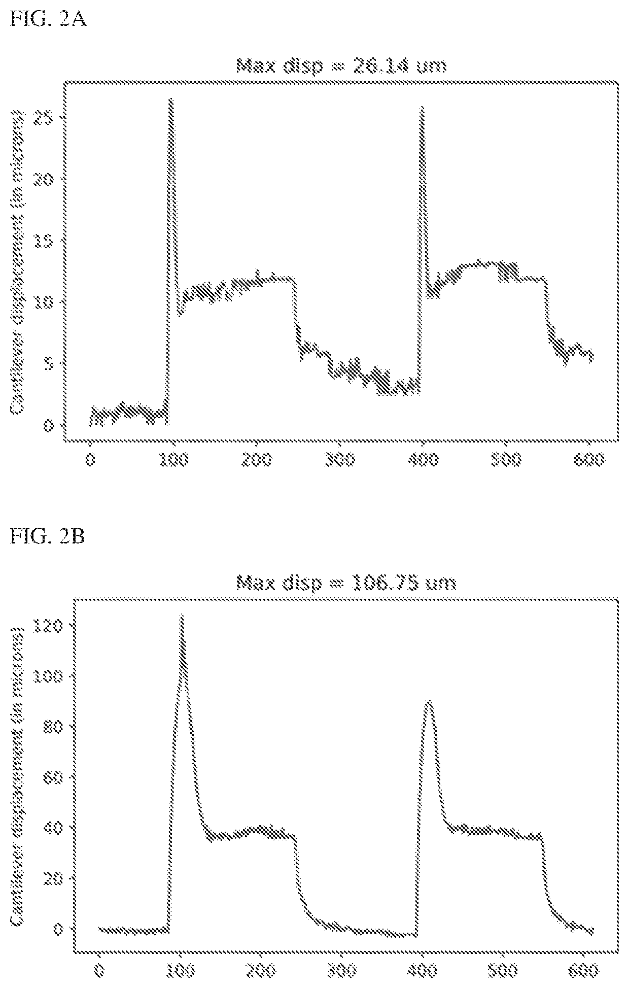
[0123]An in vitro optogenetic skeletal muscle micro-tissue as a model system (e.g., engineered skeletal muscle) such as the one shown in FIG. 1A-1E was used. Briefly, a sacrificial molding technique was used to form the muscle tissue on the cantilevers. A steel rod was used to mold a cylindrical hole in solidified gelatin. The steel rod was then removed and myoblasts in an extracellular matrix material was inserted in its place. The myoblasts were then differentiated in place to form a differentiated muscle tissue. With this system, a range of actin inhibitors were tested (e.g., Cytochalasin D (Zygosporin A), Latrunculin A, SMIFH2, CK666 and Jasplakinode) including ones that bind to the barbed end of F-actin, ones that sequester G-actin monomers and inhibitors of formins, roughly a 100% increase in muscle strength was observed (FIGS. 2A-2B and FIGS. 3A-3B) and a 300% increase in external work performed was observed, within 2 hours of F-a...
example 2
ruption Lead to a Transient Increase in Mouse Hindlimb Explant Contractility
[0131]The hindlimb extensor digitorum longus (EDL) muscle of a euthanized 2-3 month old male mouse was dissected. The EDL muscle was anchored between two kapton cantilevers using a cyanoacrylate glue. One of the cantilevers was movable whose position could be adjusted to change the resting tension of the muscle. The cantilever was moved to the point where maximal force was produced and was held at this point for the rest of the experiment. (FIG. 7A-7B). Zygosporin A (ZygoA) disrupted F-actin in the muscle explants as shown in the phalloidin stained samples after 2 hours of treatment with 3 uM ZygoA or DMSO (FIG. 7C).
[0132]Comparison of contractility changes of DMSO and ZygoA treated tissues (treatment at t=60 mins) showed a transient improvement in ZygoA treated tissues before rigor mortis leads to functional decline of the tissue. None of the DMSO treated tissues show any such transient improvement. The two...
example 3
isruption Leads to an Improvement in In Vitro Engineered Skeletal Muscle Contraction
[0133]Construction of optogenetic transgenic C2C12 myoblast cell line and Cyto D (e.g., Zygo A) treatment of differentiated C2C12 myotubes lead to a significant improvement in active contraction as quantified by changes in the Index of Movement as shown in the colormap and increased in active contractile displacement (FIG. 8A-8C). Fabrication of a 3D engineered skeletal muscle tissue by differentiating C2C12 myoblasts mixed in a fibrin / matrigel extracellular matrix cast in to a muscle-like cylindrical shape using a sacrificial molding technique. The muscle tissue is anchored to a stiff cantilever and a more compliant cantilever. Optogenetic stimulation of the muscle tissue lead to muscle contraction which was quantified through measurable displacements of the compliant cantilever. ZygoA treatment of the 3D muscle tissues lead to a significant improvement in contraction relative to DMSO treated contro...
PUM
| Property | Measurement | Unit |
|---|---|---|
| mass | aaaaa | aaaaa |
| molecular mass | aaaaa | aaaaa |
| molecular mass | aaaaa | aaaaa |
Abstract
Description
Claims
Application Information
 - R&D
- Intellectual Property
- Life Sciences
- Materials
- Tech Scout
- Unparalleled Data Quality
- Higher Quality Content
- 60% Fewer Hallucinations
Browse by: Latest US Patents, China's latest patents, Technical Efficacy Thesaurus, Application Domain, Technology Topic, Popular Technical Reports.
© 2025 PatSnap. All rights reserved.Legal|Privacy policy|Modern Slavery Act Transparency Statement|Sitemap|About US| Contact US: help@patsnap.com


