Method for recognizing medical image and system of same

a medical image and recognition technology, applied in image analysis, image enhancement, healthcare informatics, etc., can solve the problems of difficult to improve the accuracy of pre-training model medical images, and achieve the effect of improving the accuracy of medical image classification

Active Publication Date: 2021-03-25
MUEN BIOMEDICAL & OPTOELECTRONIC TECH INC
View PDF0 Cites 0 Cited by
  • Summary
  • Abstract
  • Description
  • Claims
  • Application Information

AI Technical Summary

Benefits of technology

[0001]The present disclosure relates to the field of medical image recognition, and more particularly, to a method and a system for medical image recognition applied to X-ray, computerized tomography (CT), magnetic resonance imaging (MRI), ultrasound,

Problems solved by technology

However, due to the confidentiality of personal medical information, public databases such as ImageNet lack a large number of medical images for medical image reco

Method used


Image

  • Method for recognizing medical image and system of same
  • Method for recognizing medical image and system of same
  • Method for recognizing medical image and system of same

Examples


second embodiment

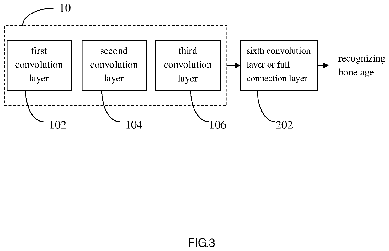
[0026]After the above pre-training is completed, the base structure 10 of this pre-trained model is applied to the image database of bone age prediction provided by RSNA to establish a model that can identify bone age. Referring to the medical image recognition system of a second embodiment shown in FIG. 3, the base structure 10 calculates and analyzes the image database of the bone age prediction provided by the RSNA through the first convolution layer 102, the second convolution layer 104 and the third convolution layer 106, the output data is passed through a sixth convolution layer (or a fully connected layer) 202 to identify the bone age. The number of convolution layers in this embodiment can be adjusted according to actual application requirements. In the practical application of this embodiment, other computing layers may also be added according to different requirements, such as a rectified linear Layer, a pooling layer, and the like. The field of image recognition applicab...

third embodiment

[0029]During the training of the pre-trained model in the third embodiment, the total number of different disease marker types used in steps 302, 304, and 306 are at least two or more types, and each image data has at least one disease marker, the number can be increased according to the needs of the actual application.

[0030]Since the features of the preceding few layers of the pre-trained model are similar under the same image recognition application scenario, the parameters of the preceding few layers can be frozen during training to improve the accuracy of image recognition. Referring to the fourth embodiment shown in FIG. 6, the fourth embodiment is a method flowchart of establishing a pre-trained model of medical images, in which parameters of at least one convolution layer in a base structure are frozen. The method flowchart of the fourth embodiment includes the following steps:

step 402: inputting image data with a D0 disease marker;

step 404: inputting image data with a D1 dis...

fourth embodiment

[0031]During the training of the pre-trained model in the fourth embodiment, the total number of different disease marker types used in steps 402, 404, and 406 are at least two or more types, and each image data has at least one disease marker, the number can be increased according to the needs of the actual application.

[0032]Referring to FIG. 7, it is experimentally confirmed that under the same image recognition application scenario, experimental analysis of different levels of magnitude is performed, the parameters of several preceding convolution layers are frozen, and only the convolution layers after the specific preceding convolution layers are trained, the prediction accuracy of the obtained pre-trained model can be increase. The improvement of the prediction accuracy is more obvious especially in the case of less data. The accuracy rate of image recognition obtained by using the pre-trained model of the present disclosure having frozen several preceding convolution layers f...


PUM


Abstract

The present disclosure provides a method and a system for recognizing medical image, the present disclosure utilizes the image data with markers of different diseases for calculating and analyzing to build a pre-trained model, the present disclosure has significant improvements to improve the accuracy of image recognition under the general situation of insufficient effective data in the field of medical image recognition technology, the present disclosure can be applied to the field of medical image recognition technology, including X-ray, CT, MRI, ultrasonic, pathological slice photography or fundus photography.

Description

BACKGROUND OF INVENTIONField of Invention[0001]The present disclosure relates to the field of medical image recognition, and more particularly, to a method and a system for medical image recognition applied to X-ray, computerized tomography (CT), magnetic resonance imaging (MRI), ultrasound, pathological slice photography or fundus photography. The method and system are used to establish a pre-trained model based on medical images to improve the accuracy of medical image classification, target detection, segmentation and other applications.Description of Prior Art[0002]In recent years, because ImageNet has provided databases of millions images, such as cats, dogs, airplanes, bicycles, and different types of object pictures, the artificial intelligence image recognition technology with deep learning neural network technology has been built to be driven by big data. The current deep learning technology of artificial intelligence can have a recognition accuracy close to human capabilit...

Claims


Application Information

Patent Timeline
IPC IPC(8): G06T7/00
CPCG06T7/0014G06T2207/20081G06T2207/10116G06T2207/20084G06T2207/10132G06T2207/30041G06T2207/10081G16H30/20G06N3/08G06N3/045G06T2207/30004
Inventor WANG, CHUEN-HENG
Owner MUEN BIOMEDICAL & OPTOELECTRONIC TECH INC
Why Eureka
Try Eureka