Distinguishing between extravascular and intravascular contrast pools

Pending Publication Date: 2021-05-27
OREGON HEALTH & SCI UNIV
View PDF0 Cites 0 Cited by
  • Summary
  • Abstract
  • Description
  • Claims
  • Application Information

AI Technical Summary

Benefits of technology

The patent is about a technique for using magnetic resonance imaging (MRI) to examine the microvasculature and cellular components of tissues, specifically by using nanoparticles as a contrast agent. This allows for the specific localization of the contrast agent within an extravascular compartment of a region of interest, where it is preferentially taken up by innate immune cells. This technique addresses a current lack of a clinically applicable biomarker for immune activity, which can be used to improve glioblastoma clinical outcomes and may also have applications for analyzing immune activity for other diseases. The disclosed technique uses noninvasive T2* weighting and susceptibility weighted sequences to specifically locate and differentiate extravascular from intravascular MRI contrast within tissues, which is important for accurate assessment of tissue microvasculature and cellular components.

Problems solved by technology

This discrepancy is likely a factor of the retrospective nature of pseudoprogression diagnosis and the lack of standardized histological criteria by which this disease process is defined.
Also, pseudoprogression cannot be distinguished from true glioblastoma regrowth using GBCA-MRI.
As such, definitive diagnosis has entailed either surgical biopsy or watchful waiting for diminishing GBCA enhancement, both of which can effectively delay definitive therapy for true disease recurrence and potentially adversely impact overall survival.
So-called early timepoint MRI (immediately following intravenous ferumoxytol administration) allows for the quantification and localization of intravascular agent; however, the prolonged circulating half-life of approximately 15 hours may limit facile interpretation of signal changes in the glioblastoma microenvironment in delayed timepoint of imaging up to 48 hours later.

Method used


Image

  • Distinguishing between extravascular and intravascular contrast pools
  • Distinguishing between extravascular and intravascular contrast pools
  • Distinguishing between extravascular and intravascular contrast pools

Examples


example experimentation

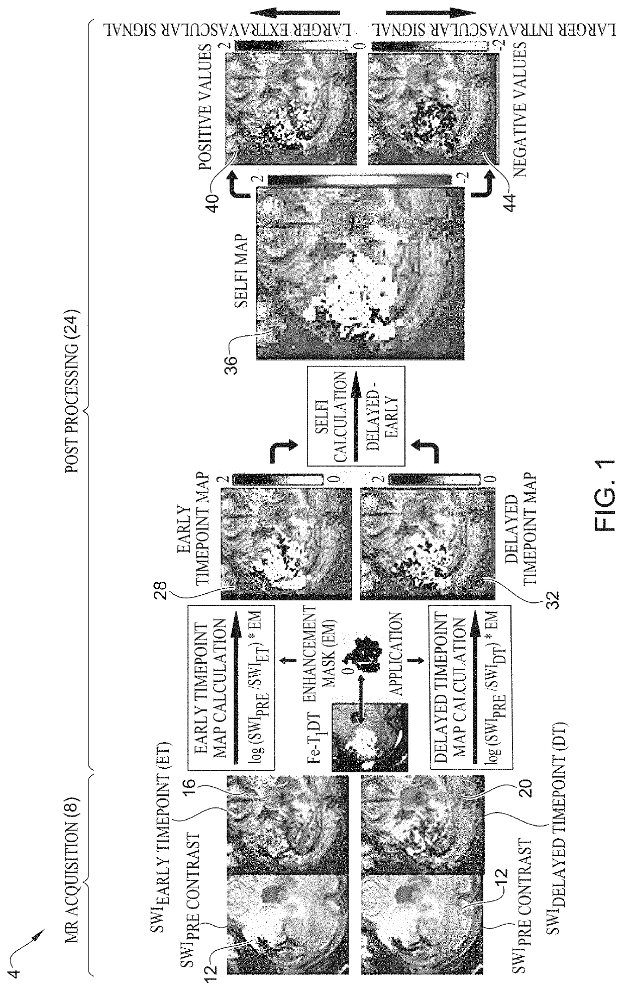
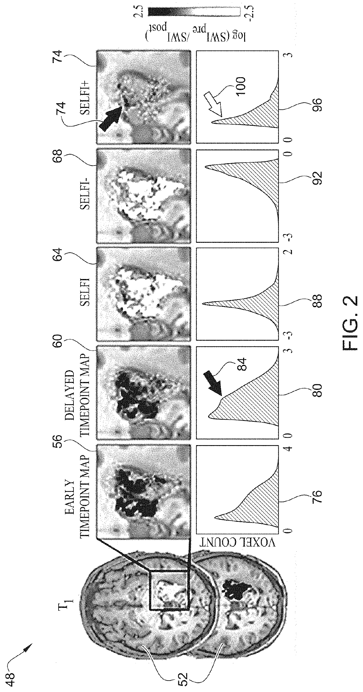
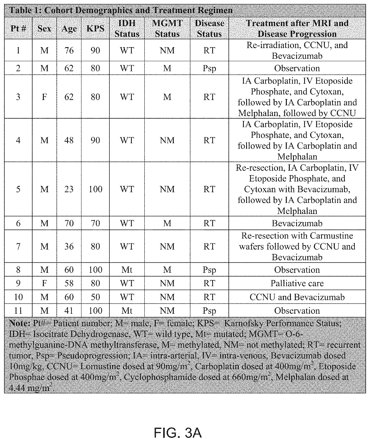
[0044]As shown in table 1 of FIGS. 3A and 3B, twenty-three patients with glioblastoma progression following Stupp protocol chemoradiotherapy were tested in an experiment. All patients underwent ferumoxytol-enhanced susceptibility weighted imaging (SWI) magnetic resonance imaging, in addition to standard T1-weighted sequences. Imaging was performed prior to, immediately after (early timepoint), and 24-hours after (delayed timepoint) intravenous ferumoxytol administration. SELFI maps were generated as the voxel-wise difference of the 24-hour delayed timepoint map from the early timepoint map. Prespecified end points included negative and positive portions of SELFI maps. Pearson's r- and Student's t-tests were performed. In terms of survival analysis, overall survival was classified in IDH wild type glioblastoma from the date of surgical diagnosis until death or last follow-up date for patients without an event. Survival analysis was performed by using multivariate Cox regression analy...


PUM


Abstract

Embodiments of the present disclosure relate to methods, apparatus, and computer readable medium for detecting an immune factor responsive to a contrast agent. In some embodiments, pre-contrast image data, early timepoint image data, and delayed timepoint image data are received. In some embodiments, an early timepoint map is generated using a comparison of the pre-contrast image data to the early timepoint image data, and a delayed timepoint map is generated using a comparison of the pre-contrast image data to the delayed timepoint image data, where the early timepoint map represents intravascular contrast, the delayed timepoint map represents the intravascular contrast and extravascular contrast, and the extravascular contrast indicates the immune factor. In some embodiments, a combined immune factor map is generated by voxel-wise subtraction of the early timepoint map from the delayed timepoint map.

Description

RELATED APPLICATION[0001]This application claims priority benefit of U.S. Provisional Patent Application No. 62 / 938,789, filed Nov. 21, 2019, which is hereby incorporated by reference in its entirety.FEDERALLY SPONSORED RESEARCH OR DEVELOPMENT[0002]This invention was made with government support under R01 CA137488 awarded by the National Institutes of Health. The government has certain rights in the invention.TECHNICAL FIELD[0003]This disclosure relates generally to analysis of magnetic resonance imaging (MRI) images and, more particularly, to detection of an immune factor.BACKGROUND INFORMATION[0004]Despite advances in surgical resection and Stupp protocol adjuvant chemoradiotherapy (e.g., CRT; temozolomide with conformal irradiation), the median overall survival is currently about 14.2 months in isocitrate dehydrogenase (IDH) wild-type glioblastoma. Gadolinium-based contrast (GBCA) enhanced magnetic resonance imaging (MRI) is a technique employed for therapeutic monitoring of glio...

Claims


Application Information

Patent Timeline
IPC IPC(8): G01R33/56G06T7/00G16H30/40G16H50/20A61B5/00A61B5/055G01R33/50
CPCG01R33/5601G06T7/0016G16H30/40G16H50/20A61B5/0042A61B2576/026G01R33/50G06T2207/30104G06T2207/30096G06T2207/10088G06T2207/20224A61B5/055G06T2207/20016G06T2207/30016G06T7/32G16H20/17G01R33/5602A61B5/41A61B5/4064
Inventor BARAJAS, JR., RAMON FRANCISCOSCHWARTZ, DANIEL
Owner OREGON HEALTH & SCI UNIV
Why Eureka
Try Eureka