
Short t2 tissue imaging with t2 prep petra sequence

a tissue imaging and short t2 technology, applied in the field of magnetic resonance imaging, can solve the problem of limited suppression of long t2 tissu

Pending Publication Date: 2021-06-10
SIEMENS HEALTHCARE GMBH
View PDF2 Cites 0 Cited by
  • Summary
  • Abstract
  • Description
  • Claims
  • Application Information

AI Technical Summary

Benefits of technology

The invention is about improving magnetic resonance imaging (MRI) using a technique called PETRA. In this method, a specific pulse sequence is used to capture images of different tissues in the body. By combining two different pulse sequences, the method allows for better imaging of certain tissues and reduces the time needed for the MRI scan. Additionally, a technique called a spoiled gradient is used to improve image quality. This invention expands the application range of the PETRA technique and makes it more patient-friendly. Overall, this patent improves the efficiency and accuracy of MRI scans.

Problems solved by technology

Although the single-echo PETRA technique can acquire an image of a short T2 tissue, it has limited suppression of the long T2 tissue.

Method used


Image

  • Short t2 tissue imaging with t2 prep petra sequence
  • Short t2 tissue imaging with t2 prep petra sequence
  • Short t2 tissue imaging with t2 prep petra sequence

Examples


Embodiment Construction

[0014]In the following description, numerous specific details are set forth in order to provide a thorough understanding of the embodiments of the present disclosure. However, it will be apparent to those skilled in the art that the embodiments, including structures, systems, and methods, may be practiced without these specific details. The description and representation herein are the common means used by those experienced or skilled in the art to most effectively convey the substance of their work to others skilled in the art. In other instances, well-known methods, procedures, components, and circuitry have not been described in detail to avoid unnecessarily obscuring embodiments of the disclosure. The connections shown in the figures between functional units or other elements can also be implemented as indirect connections, wherein a connection can be wireless or wired. Functional units can be implemented as hardware, software or a combination of hardware and software.

[0015]An a...


PUM


Abstract

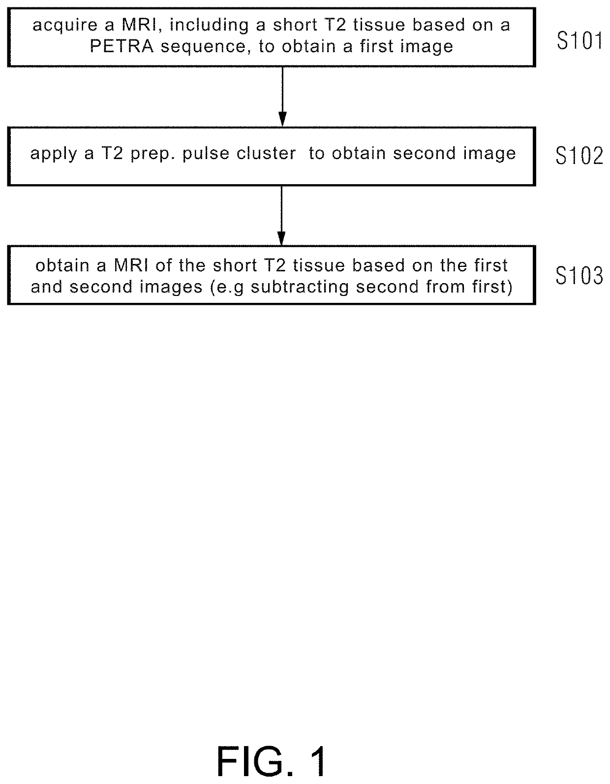
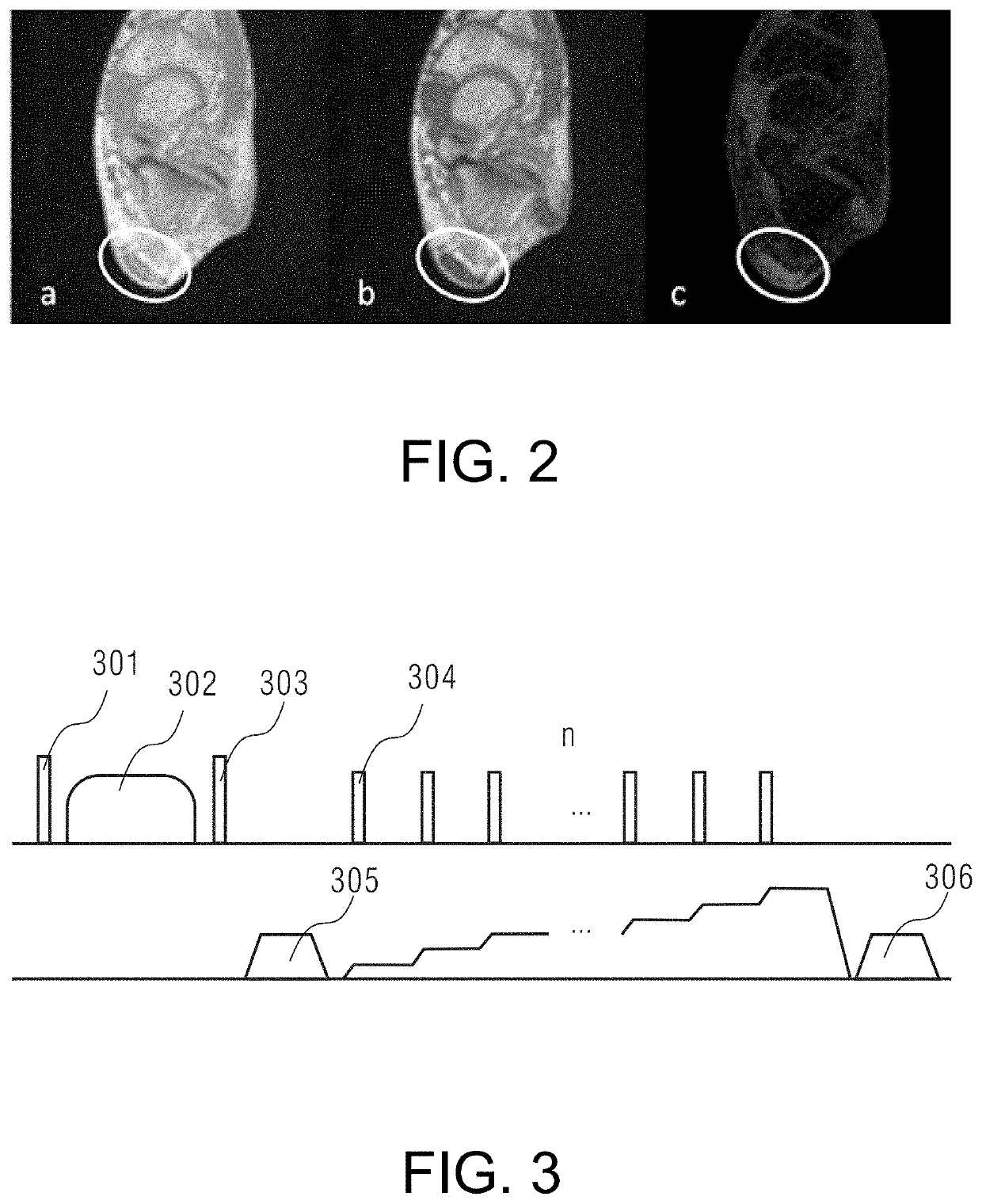
In a short T2 tissue imaging method and system, a magnetic resonance image is acquired that includes a short T2 tissue based on point-wise encoding time reduction with radial acquisition point-wise encoding time reduction with radial acquisition (PETRA) sequences, to obtain a first image, a T2 preparation pulse cluster is applied for suppressing a short T2 tissue signal between the PETRA sequences according to a predetermined interval of applying the T2 preparation pulse cluster, a magnetic resonance image is acquired that excludes the short T2 tissue based on the PETRA sequences applied with the T2 preparation pulse cluster, to obtain a second image; and a magnetic resonance image of the short T2 tissue is obtained based on the second image and the first image (e.g. by subtracting the second image from the first image).

Description

CROSS REFERENCE TO RELATED APPLICATIONS[0001]This patent application claims priority to Chinese Patent Application No. 201911258002.X, filed Dec. 10, 2019, which is incorporated herein by reference in its entirety.BACKGROUNDField[0002]The present disclosure relates to the field of magnetic resonance imaging, and in particular to a short T2 tissue imaging method, a short T2 tissue imaging system, and a magnetic resonance imaging system.Related Art[0003]Magnetic resonance imaging is an imaging technique using a magnetic resonance phenomenon. The principle of magnetic resonance phenomenon mainly involves nuclei containing an odd number of protons, for example, hydrogen nuclei widely existing in a human body, the protons thereof being in a spin motion, like small magnets, and the small magnets having irregular axes of spin. If an external magnetic field is applied, the small magnets will be rearranged according to magnetic lines of force of the external magnetic field, and are specifica...

Claims


Application Information

Patent Timeline
IPC IPC(8): G01R33/561G01R33/50A61B5/055
CPCG01R33/561A61B5/055G01R33/50G01R33/5616A61B5/6829A61B5/4523G01R33/5602G01R33/4824
Inventor DONG, FANGZHOU, KUN
Owner SIEMENS HEALTHCARE GMBH
Learn More