Combinatorial use of markers to isolate synaptic glia to generate synapses in a dish for high-throughput and high-content drug discovery and testing

a synapses and marker technology, applied in the chemical analysis of biological materials, instruments, material analysis, etc., can solve the problem of slow progress in answering fundamental questions about synaptic glia

Pending Publication Date: 2021-11-11
BROWN UNIVERSITY
View PDF0 Cites 2 Cited by
  • Summary
  • Abstract
  • Description
  • Claims
  • Application Information

AI Technical Summary

Benefits of technology

[0020]In another aspect, the invention provides a way to understand how the three cellular constituents of the synapse—neurons, muscle, and glia—communicate each other. The invention provides a tool,

Problems solved by technology

The slow progress in answering fundamental questions about synaptic glia can is primarily du

Method used


Image

  • Combinatorial use of markers to isolate synaptic glia to generate synapses in a dish for high-throughput and high-content drug discovery and testing
  • Combinatorial use of markers to isolate synaptic glia to generate synapses in a dish for high-throughput and high-content drug discovery and testing
  • Combinatorial use of markers to isolate synaptic glia to generate synapses in a dish for high-throughput and high-content drug discovery and testing

Examples


example 1

Identification of a Molecular Fingerprint for Synaptic Glia

[0068]The inventors explored the possibility that synaptic glia can be distinguished by unique combinations of glial cell markers, determined by a cell-specific pattern of gene expression. Synaptic glia of both the central (CNS) and peripheral (PNS) nervous systems are generally thought in the biomedical art to provide structural, functional, and trophic support to the synapse. The inability to selectively visualize and target perisynaptic Schwann cells remains an obstacle to understanding the cellular and molecular rules that govern their differentiation and function at neuromuscular junctions during development, following injury, in old age, and diseases, such as ALS.

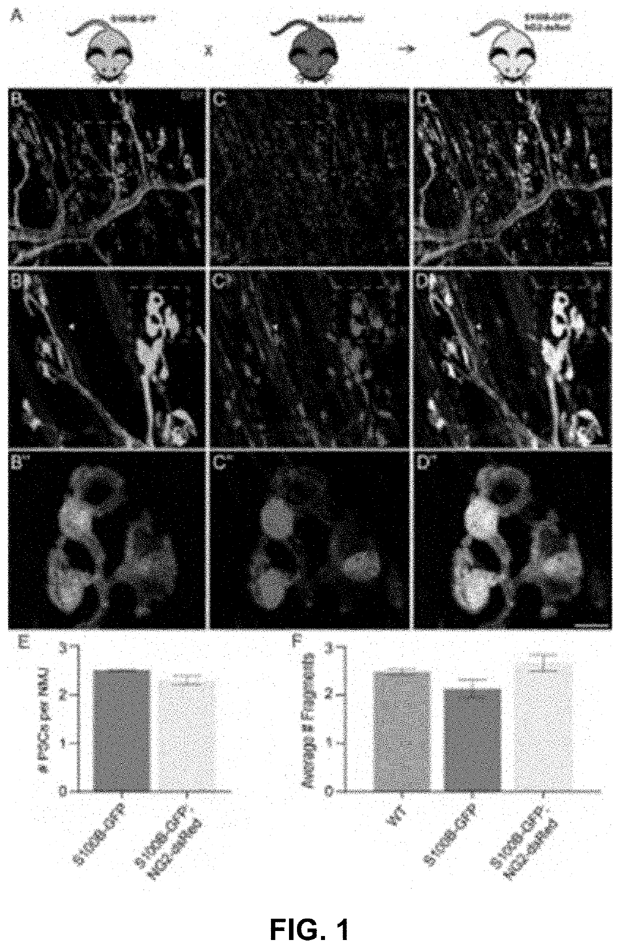
[0069]To facilitate visualization of perisynaptic Schwann cells, the inventors created a transgenic mouse line (called S100β-GFP;NG2-dsRed; see FIG. 1(A)) by crossing transgenic lines in which either the NG2 promoter, which drives expression of dsRed; see Zhu,...

example 2

The S100β-GFP;NG2-dsRed Mouse Line is a Reliable Model to Study Perisynaptic Schwann Cells

[0081]The inventors evaluated whether the S100β-GFP;NG2-dsRed mouse line is a reliable model to study perisynaptic Schwann cells and their functions at neuromuscular junctions. In healthy young adult muscle, the inventors observed the same number of perisynaptic Schwann cells at neuromuscular junctions in the extensor digitorum longus muscle of S100β-GFP and S100β-GFP;NG2-dsRed mice. See FIG. 1(E). The morphology of perisynaptic Schwann cells also appeared to be indistinguishable between the two transgenic lines. The morphology of neuromuscular junctions, as assessed by fragmentation of nicotinic acetylcholine receptor (nAChR) clusters, is unchanged in S100β-GFP;NG2-dsRed mice compared to S100β-GFP and wild type mice. See FIG. 1(F). Thus, the coëxpression of S100β-GFP and NG2-dsRed does not appear to cause apparent deleterious changes on either perisynaptic Schwann cells or the postsynaptic reg...

example 3

The Relationship Between NG2 Expression and Perisynaptic Schwann Cell Differentiation

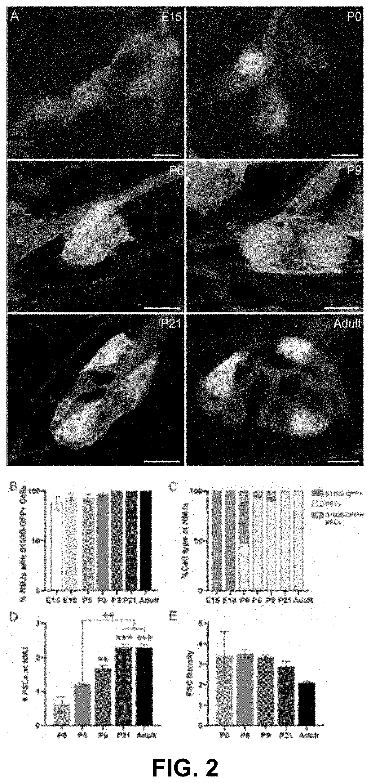
[0085]The inventors analyzed NG2 expression in S100β-GFP+ Schwann cells during the course of neuromuscular junction development in the extensor digitorum longus muscle of S100β-GFP;NG2-dsRed mice. See FIG. 2(A). The inventors observed the presence of S100β-GFP+ cells at the neuromuscular junction as early as embryonic day 15 (E15) with 100% of neuromuscular junctions having at least one S100β-GFP+ cell by post-natal day 9. See FIG. 2(A)-(B). During the embryonic developmental stages, neuromuscular junctions are exclusively populated by S100β-GFP+ cells that do not express NG2-dsRed. See FIG. 2(C). At post-natal day 0 (P0), however, NG2-dsRed expression in a small subset of S100β-GFP+ cells. See FIG. 2(A)&(C). Surprisingly, the proportion of neuromuscular junctions with S100β-GFP+;NG2-dsRed+ cells sharply increased between the ages of P0 and P9, coinciding with the period of neuromuscular junction ma...


PUM


Abstract

The invention provides molecular tools to visualize, isolate, and manipulate the glial cells that are necessary for the formation, stability, and function of the synapse. The inventors identified a unique gene expression signature that distinguishes perisynaptic Schwann cells from all other Schwann cells. Using a combinatorial approach and coëxpressing two different fluorescence proteins, each using a different promoter, only those glial cells associated with the neuromuscular synapse are labeled.

Description

REFERENCE TO RELATED APPLICATIONS[0001]This invention claims priority under 35 U.S.C. 119(e) to the provisional patent application U.S. Ser. No. 63 / 013,344, titled “Combinatorial use of markers to isolate synaptic glia to generate synapses in a dish for high-throughput and high-content drug discovery and testing” and filed on Apr. 21, 2020, the entire contents of which are hereby incorporated herein by reference.STATEMENT REGARDING FEDERALLY SPONSORED RESEARCH OR DEVELOPMENT[0002]This invention was made with government support under Grant Numbers R01 AG055545 and R21 NS106313 awarded by the National Institutes of Health. The government has certain rights in the invention.FIELD OF THE INVENTION[0003]This invention generally relates to the chemical analysis of biological material, including the testing involving biospecific ligand binding methods, such as immunological testing, the measuring or testing processes involving enzymes or microorganisms, compositions or test papers, process...

Claims


Application Information

Patent Timeline
IPC IPC(8): G01N33/50G01N33/68G01N15/14
CPCG01N33/5058G01N33/68G01N2015/0065G01N2333/705G01N2333/43595G01N15/1434G01N15/1475G01N2015/149G01N15/1459G01N2015/1006G01N2015/1488
Inventor VALDEZ, GREGORIO
Owner BROWN UNIVERSITY
Why Eureka
Try Eureka