Specimen analysis method and image processing method

a technology of image processing and specimen analysis, applied in image enhancement, image data processing, instruments, etc., can solve the problems of difficult to clearly separate individual cells in specimens in which cells exist at a high density, loss of cell position information in specimens, and inability to apply a technique immediately to multiple immunostained specimens

Pending Publication Date: 2022-03-03
DAINIPPON SCREEN MTG CO LTD +1
View PDF0 Cites 0 Cited by
  • Summary
  • Abstract
  • Description
  • Claims
  • Application Information

AI Technical Summary

Benefits of technology

The invention provides a method for extracting individual cells from a high-density tissue specimen image using image processing techniques. The method accurately specifies the boundaries of each cells by extracting the inner regions inward of the cell membranes and expanding them outward. This allows for the quantitative analysis of each cell in the tissue specimen and the extraction of individual pieces of quantitative information.

Problems solved by technology

This flow cytometry technique has a problem that the position information of cells in a specimen is lost.
That is, a method for specifying the positions of the biological materials is not described in detail in patent literature 1, but it is possibly difficult to clearly separate individual cells in a specimen in which the cells exist at a high density.
However, since the above conventional technique has constraint conditions for staining methods, this technique cannot be immediately applied to such a multiple immunostained specimen.
In such a tissue section, it is difficult to separate individual cells in an image since the cells overlap also in a depth direction.
This problem is particularly prominent in a specimen having a high cell density.
If the tissue section is thinned, it is advantageous in terms of cell discrimination, but the number of times of staining is limited due to another problem that the specimen is easily damaged.

Method used


Image

  • Specimen analysis method and image processing method
  • Specimen analysis method and image processing method
  • Specimen analysis method and image processing method

Examples


Embodiment Construction

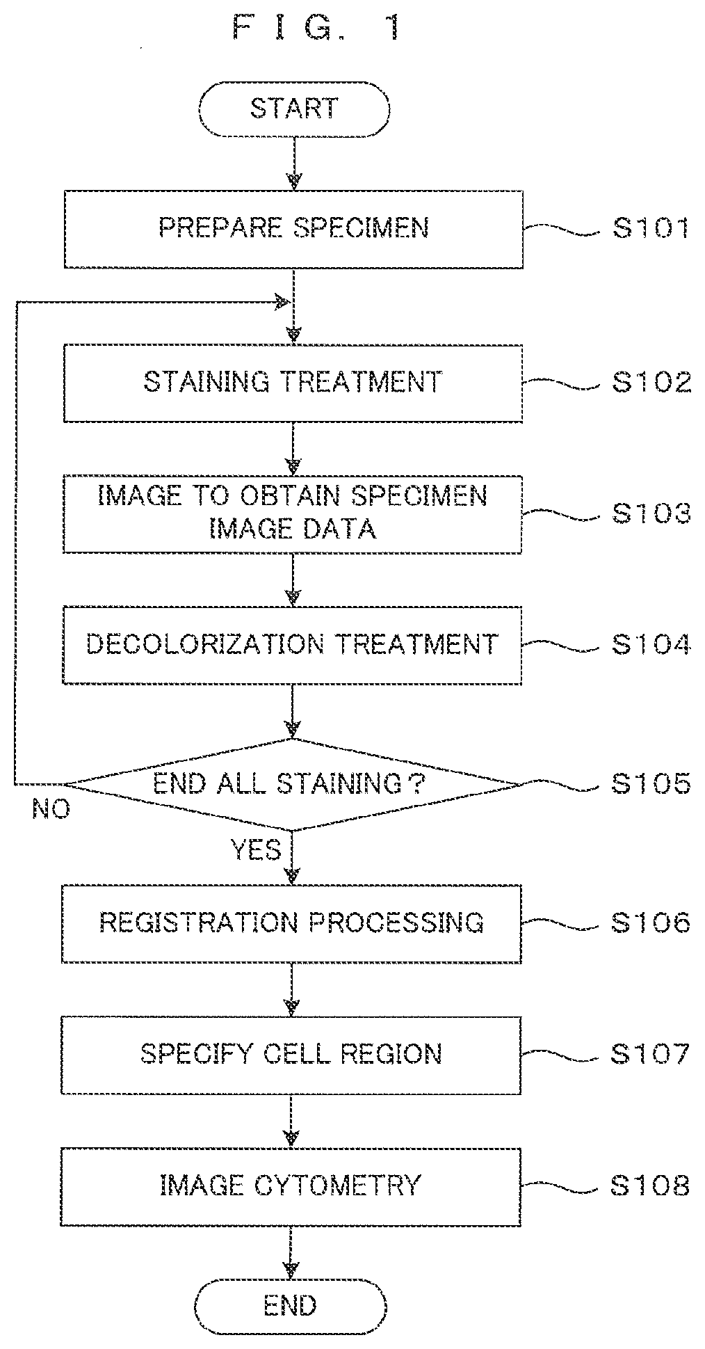
[0024]Hereinafter, a specimen analysis process (hereinafter, merely referred to as an “analysis process”), which is one embodiment of a specimen analysis method according to the invention, is described. This analysis process is a process for specifying the positions of individual cells from an image of a specimen including a multitude of cells such as a pathological tissue specimen and obtaining quantitative information such as expression states and expression levels of biological materials in each cell. A basic technical concept of this process follows a multiple immunostaining technique and is not described since a basic principle of multiple immunostaining is known. First, this analysis process is summarized with reference to FIG. 1 and, then, the content of each processing step is described in detail.

[0025]FIG. 1 is a flow chart showing a flow of the specimen analysis process of this embodiment. First, a pathological tissue specimen to be analyzed is prepared (Step S101). For ex...


PUM


Abstract

An image processing using a plurality of specimen images obtained by successively applying a plurality of types of staining to a specimen to be evaluated and imaging the specimen after staining for at least two types of staining is performed. The image processing comprises: extracting an inner region corresponding to inward of a cell membrane of a single cell in the specimen based on at least one of the specimen images, specifying a cell region corresponding to an individual cell included in the specimen by expanding the inner region outwardly, and performing image cytometry for the cell region based on the specimen images. In analyzing a pathological specimen on a cell-by-cell basis, it is possible to deal with multiple immunostaining and specify the positions of individual cells and evaluate each cell separately even when a cell density in the specimen is high.

Description

CROSS REFERENCE TO RELATED APPLICATION[0001]The disclosure of Japanese Patent Application No. 2020-141765 filed or Aug. 25, 2020 including specification, drawings and claims is incorporated herein by reference in its entirety.BACKGROUND OF THE INVENTION1. Field of the Invention[0002]This invention relates to a method for analyzing a specimen image captured by imaging a pathological specimen on a cell-by-cell basis and an image processing method for assisting the former method, particularly to an analysis using an immunohistochemical technique.2. Description of the Related Art[0003]In the field of recent immunohistochemistry, a so-called single cell analysis not only seeing a specimen as a group of cells, but also focused on individual cells constituting the specimen is attracting attention. This is a method for measuring expression levels, positions and the like of biological materials such as proteins and protein-related materials on a cell-by-cell basis. A flow cytometry technique...

Claims


Application Information

Patent Timeline
IPC IPC(8): G06T7/00G06K9/20G06T7/30G06T7/187G01N15/14
CPCG06T7/0014G06K9/2054G06T7/30G01N2015/1006G01N15/1429G06T2207/30024G06T2207/10056G06T7/187G06T7/11G06T2207/10024G06T2207/10016G06T2207/20081G06T7/0012G06T2207/10064G01N33/4833G06V10/22G01N2015/1497G01N15/01
Inventor HIRAI, MAKIOGI, HIROSHIISHIHARA, SHUNTATSUJIKAWA, TAKAHIROITOH, KYOKO
Owner DAINIPPON SCREEN MTG CO LTD
Why Eureka
Try Eureka