Systems and Methods for Generating Quick-Glance Interactive Diagnostic Reports

Pending Publication Date: 2022-03-17
DIAGNOCAT INC
View PDF0 Cites 3 Cited by
  • Summary
  • Abstract
  • Description
  • Claims
  • Application Information

AI Technical Summary

Benefits of technology

This patent describes a system that uses computer software and hardware to perform medical imaging tasks. The system includes a system for analyzing and categorizing dental conditions from images. Another part of the system helps create a visual representation of the dental condition. The patent also describes the use of augmented reality to assist dental professionals in diagnosing and treating tooth issues. The overall technical effects of this patent are improved automation and efficiency in handling dental imaging tasks, leading to improved dental outcomes.

Problems solved by technology

However, CBCT includes one or more limitations, such as time consumption and complexity for personnel to become fully acquainted with the imaging software and correctly using digital imaging and communications in medicine (DICOM) data.
Further, many dental professionals who incorporate this technology into their practices have not had the training required to interpret data on anatomic areas beyond the maxilla and the mandible.
To address the foregoing issues, deep learning has been applied to various medical imaging problems to interpret the generated images, but its use remains limited within the field of dental radiography.
Further, most applications only work with 2D X-ray images.
However, this method fails to be sufficiently robust for practical use.
However, the above references also fail to address the afore discussed problems regarding the cone beam computed tomography technology and image generation system, not to mention an automated anatomical localization and pathology detection / classification means.
The extant annotation tools do not support localizing and enumerating teeth in the images using neural networks, and certainly, do not support sorting images into a full mount table with neural network-mediated classification of a tooth condition with diagnostic value.
Therefore, there is void in the art for an automated localization, numeration, and diagnostic system and method for FMX and panoramic images for improved dental health outcomes.

Method used


Image

  • Systems and Methods for Generating Quick-Glance Interactive Diagnostic Reports
  • Systems and Methods for Generating Quick-Glance Interactive Diagnostic Reports
  • Systems and Methods for Generating Quick-Glance Interactive Diagnostic Reports

Examples


Embodiment Construction

[0059]Specific embodiments of the invention will now be described in detail with reference to the accompanying FIGS. 1A-7. In the following detailed description of embodiments of the invention, numerous specific details are set forth in order to provide a more thorough understanding of the invention. In other instances, well-known features have not been described in detail to avoid obscuring the invention. Embodiments disclosed include an automated parsing pipeline system and method for anatomical localization and condition classification.

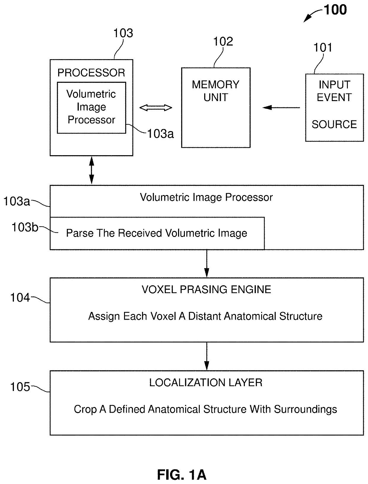
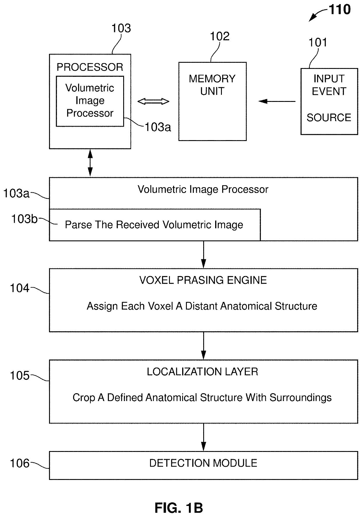
[0060]FIG. 1A illustrates a block diagram 100 of the system comprising an input event source 101, a memory unit 102 in communication with the input event source 101, a processor 103 in communication with the memory unit 102, a volumetric image processor 103a in communication with the processor 103, a voxel parsing engine 104 in communication with the volumetric image processor 103a and a localizing layer 105 in communication with the voxel parsing ...


PUM


Abstract

A system for an infographic display of an image analysis, said system comprising: a processor; a non-transitory storage element coupled to the processor; encoded instructions stored in the non-transitory storage element, wherein the encoded instructions when implemented by the processor, configure the system to: receive at least one image frame; localize at least one of a present tooth, dental, or non-dental condition inside the received image frame and identify it by at least one of a number, name, or short-hand; extract the at least one identified tooth, dental, or non-dental condition within the received image; classify the at least one tooth, dental, or non-dental condition based on the extracted; and represent results of the at least one classified area of interest in at least one of three layers, wherein the layers are an image-based layer, infographic-based layer, or an informational layer.

Description

BACKGROUNDField[0001]This invention relates generally to medical diagnostics, and more specifically, to an automated tooth localization, enumeration, and diagnostic tool featuring visuals and cursor control mechanisms for enabling quick-glance analysis of various diagnostic layers.RELATED ART[0002]Modern image generation systems play an important role in disease detection and treatment planning. Few existing systems and methods were discussed as follows. One common method utilized is dental radiography, which provides dental radiographic images that enable the dental professional to identify many conditions that may otherwise go undetected and to see conditions that cannot be identified clinically. Another technology is cone beam computed tomography (CBCT) that allows to view structures in the oral-maxillofacial complex in three dimensions. Hence, cone beam computed tomography technology is most desired over the dental radiography.[0003]However, CBCT includes one or more limitations...

Claims


Application Information

Patent Timeline
IPC IPC(8): G06T11/60G06K9/62G06T7/00G06T7/10G06V10/82A61B6/14A61B6/03A61B6/00
CPCG06T11/60G06K9/6268G06T7/0012G06T7/10G06V10/82A61B6/14G06T2207/10081A61B6/4085A61B6/463G06T2207/30036G06T2207/20084G06T2200/24G06T2207/20081A61B6/032A61B5/0022A61B5/1075A61B5/4504A61B5/4842A61B5/7267A61B5/7275A61B5/743A61B6/501G06N3/084G06V10/255G06V2201/03G16H30/20G16H50/20G16H30/40G16H15/00G16H50/70G06N3/082G06N3/048G06N3/044G06N3/045A61B6/51G06F18/241
Inventor EZHOV, MATVEY DMITRIEVICHALEKSANDROVSKIY, VLADIMIR LEONIDOVICHSHUMILOV, EVGENY SERGEEVICH
Owner DIAGNOCAT INC
Why Eureka
Try Eureka