Comprehensive detection of single cell genetic structural variations

a single cell, genetic structural technology, applied in the field of comprehensive detection of single cell genetic structural variations, can solve the problems of limiting the utility of sv detection in heterogeneous contexts, sv discovery remains challenging, and svs represent a particularly difficult-to-identify class of variation, so as to achieve a different diagnostic footprint

Pending Publication Date: 2022-06-23
MAX PLANCK GESELLSCHAFT ZUR FOERDERUNG DER WISSENSCHAFTEN EV +2
View PDF0 Cites 0 Cited by
  • Summary
  • Abstract
  • Description
  • Claims
  • Application Information

AI Technical Summary

Benefits of technology

[0040]For purposes of the present invention, the term “copy-number variations (CNVs)” refers to a form of structural variation of the DNA of a genome that results in the cell having an abnormal or, for certain genes, a normal variation in the number of copies of one or more sections of the DNA. CNVs correspond to relatively large regions of the genome that have been deleted (fewer than the normal number) or duplicated (more than the normal number) on certain chromosomes. Correspondingly, the term “copy number neutral” shall denote a variation that does not result in the cell having unusual copy numbers of sequence elements such as genes.
[0041]The term “diagnostic footprint” in context of the present invention shall mean a pattern of the three layered information of the invention that is specific or at least indicative for a SV. A diagnostic footprint is therefore characterized by an alteration of the data distribution expected for a specific experiment. The specific pattern that indicates a SV will vary depending on the analysed data. For example a diploid cell may be sequenced to contain for each chromosome a WW, CC or WC strand distribution. Depending on the strand distribution, the same SV may have a different diagnostic footprint. Such footprints or patterns are for example provided herein in table 1.
[0042]In context of the herein disclosed invention the term “target chromosomal region” shall refer to a DNA sequence of one or more, full or partial, chromosomes of any organism or virus, which is the object of an inquiry in context of the invention. A target chromosomal region may refer to just one sequence of a part of a single chromosome, or to both the paternal and maternal region of any chromosome. In some embodiments the target chromosomal region which is the object of an inquiry according to the invention is a whole chromosome or a whole genome of a single cell, or a plurality of a single cell.
[0043]In context of the herein disclosed invention the term “single cell” shall refer to one individual cell from which by for example strand-specific sequencing, a single cell library is generated. A single cell library in context of the invention describes the plurality of sequence reads obtained by sequencing the genome of said single cell. Furthermore, the invention in some aspects and embodiments refers to a plurality of single cells, or multiplicity of single cells, which in this case refers to the generation of a plurality of separate and independent sequence libraries for each single cell contained in the plurality of single cells. In one preferred embodiment of the invention, up to 96 single cells of a cell line are sequenced individually. Such embodiments are preferred as such assays can be performed in multiwall plates such as 96 well plates or 384 well plates.
[0044]The term “reference sequence of the at least one target chromosomal region” refers to a database version of a fully sequenced reference of the target. Usually, such reference will be a full chromosome sequence. In some instances the reference sequence is also denoted as “reference scaffold” or “reference genomic scaffold” or “reference assembly” or similar expression. For human sequences for example the The Genome Reference Consortium frequently publishes and updates the reference sequence of the human genome, as well as other genomes such as mouse, zebrafish and chicken genomes (https: / / www.ncbi.nlm.nih.gov / grc).
[0045]The term “reference state” in context of the present invention shall refer to state or distribution of sequencing data that is used as a reference for a comparison with a sample dataset, for example in order to identify aberrations. Such a reference state may be a real set of sequencing data used as a reference, or may be state of the data that is expected for a certain underlying sampled chromosomal region. Usually a reference state in context of the invention shall pertain to the distribution of sequences within a chromosome, or set of chromosomes (genome), that is expected for a non-aberrant single cell or population of cells. As an example, a reference state of a usual diploid human genome would be a distribution of human chromosomes in somatic cells that is common to a majority of humans. However, in certain aspects and embodiments, the reference state may also contain unusual chromosomal architectures or aneuploidies—the reference state according to the invention is determined based on the samples analysed and questions to be answered with the methods of the invention. As a mere illustrative example, the sample analysed with the method of the invention may be derived from a trisomy 21 individually who is screened for other SVs. Most importantly the term “reference state” in context of the invention shall not be confused with “reference sequence”, the latter being defined above and referring to an assembly of sequences that is used for aligning sequence reads.

Problems solved by technology

SV discovery however remains challenging, with translocations, inversions, complex SV classes, cellular ploidy alterations and SVs arising in repetitive regions frequently escaping detection in genetic heterogeneity contexts.
Whether arising in the germline or somatically, SVs represent a particularly difficult-to-identify class of variation.
These methods require extensive sequence coverage for confident SV calling (˜20-fold or higher when bulk sequencing is used)17, which limits their utility for SV detection in heterogeneous contexts—with the exception of read-depth analysis, which can be pursued for variants with relatively low VAF (typically 10% VAF), but which is limited to CNAs10.
However, while CNAs are already routinely analyzed in single cells, and scalable16 as well as commercial applications (e.g., the 10× Genomics “The Chromium Single Cell CNV Solution”) are becoming available, the detection of additional SV classes such as balanced and complex SVs in single cells faces important challenges: Currently available SV detection methodology requires the identification of reads (or read pairs) traversing the SV's breakpoints55; this remains challenging due to high coverage requirements of such approach, and low as well as uneven coverage levels including localised allelic drop outs in single cells17.
And while recent studies have shown that chimera filtering is feasible in conjunction with sufficient sequence coverage19,20, SV discovery in hundreds (or thousands) of single cells would necessitate vast sequencing costs, and accordingly has not been pursued yet.
Additionally, most current methods do not indicate which haplotype a given variant resides on, which may lead to reduced calling power compared to haplotype-aware single cell analyses57.

Method used


Image

  • Comprehensive detection of single cell genetic structural variations
  • Comprehensive detection of single cell genetic structural variations
  • Comprehensive detection of single cell genetic structural variations

Examples


example 1

ables Systematic Discovery of a Wide Variety of SV Classes in Single Cells

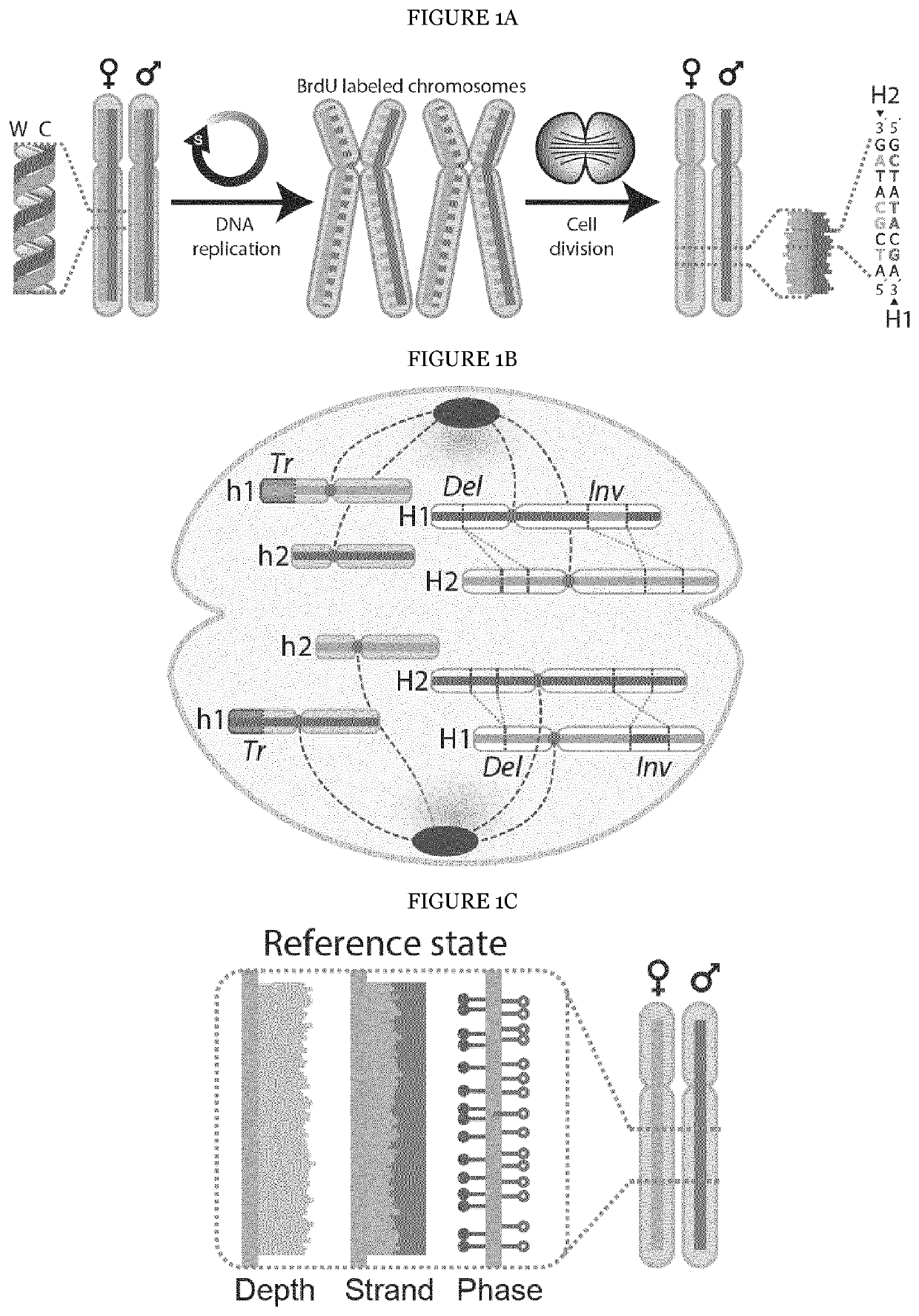
[0202]The underlying rationale of scTRIP is that each class of SV can be identified via a specific ‘diagnostic footprint’. These diagnostic footprints capture the co-segregation patterns of rearranged DNA segments made visible by sequencing single strands of each chromosome in a cell, as follows: During S-phase, the DNA double strand unwinds, and the two resulting single strands (Watson [‘W’] and Crick [‘C’]) act as templates for DNA replication. In Strand-seq, newly replicated strands incorporate Bromodeoxyuridine (BrdU)21, which acts as a traceable label for these non-template strands (see FIG. 1A depicting the Strand-seq protocol)24. During mitosis, each of the two daughter cells receive one copy of each chromosomal homolog through independent and random chromatid segregation21. The labeled nascent strand is then removed, and the segregation pattern of each chromosomal segment is analyzed following strand-s...

example 2

apes of RPE Cells Uncovered by scTRIP

[0232]To investigate single cell SV landscapes using scTRIP the inventors next generated strand-specific DNA sequencing libraries from telomerase-immortalized retinal pigment epithelial (RPE) cells. hTERT RPE cells (RPE-1) are commonly used to study patterns of genomic instability20,27-29, and additionally C7 RPE cells were used, which show anchorage-independent growth used as an indicator for cellular transformation30. Both RPE-1 and C7 cells originate from the same anonymous female donor. The inventors sequenced 80 and 154 single cells for RPE-1 and C7, respectively, to a median depth of 387,000 mapped non-duplicate fragments (Methods). This amounts to only 0.01× genomic coverage per cell.

[0233]The inventors first searched for Dels, Dups, Invs and InvDups. Following read normalization, 54 SVs in RPE-1 were identified, and 53 in C7 cells. 22 SVs were present only in RPE-1, and 21 were present only in C7, and thus likely correspond to sample-spec...

example 3

g Complex Cancer-Related Translocations in Single Cells

[0234]To assess the ability of scTRIP to detect a wider diversity of SV classes, the inventors subjected RPE-1 cells to the CAST protocol28: the inventors silenced the mitotic spindle machinery to construct an anchorage-independent line (BM510) likely to exhibit genome instability. The inventors sequenced 145 single BM510 cells detecting overall 67 SVs when searching for Dels, Dups, Invs and InvDups events. Additionally, several DNA segments did not segregate with the respective chromosomes they originated from, indicating inter-chromosomal SV formation (FIG. 3A). The inventors performed translocation detection with scTRIP searching for diagnostic co-segregation footprints (FIG. 3B), and identified four translocations in BM510 (FIG. 3B,C). The inventors additionally subjected RPE-1 and C7 to translocation detection, identifying one translocation each (FIG. 3D).

[0235]One translocation was shared between RPE-1 and BM510 and involv...


PUM


Abstract

The present invention provides a method for detecting structural variations (SV) within genomes of single cells or population of single cells by integrating a three-layered information of sequencing read depth, read strand orientation and haplotype phase. The method of the invention can detect deletions, duplications, polyploidies, translocations, inversions, and copy number neutral loss of heterozygosity (CNN-LOH), and more. The method of the invention can fully karyotype a genome comprehensively, and may be applied in research and clinical approaches. For example, the methods of the invention are useful for analysing cellular samples of patients for diagnosing or aiding a diagnosis, in reproductive medicine to detect embryonic abnormalities, or during therapeutic approaches based on cellular therapies to quality control genetically engineered cells, such as in adoptive T cell therapy and others. The method of the invention may further be applied in research to decipher the karyotypes of cellular models (cell lines), patient samples, or to further unravel genetic and mechanistic pathways leading to the generation of any SV within genomes.

Description

FIELD OF THE INVENTION[0001]The present invention provides a method for detecting structural variations (SV) within genomes of single cells or population of single cells by integrating a three-layered information of sequencing read depth, read strand orientation and haplotype phase. The method of the invention can detect deletions, duplications, polyploidies, translocations, inversions, and copy number neutral loss of heterozygosity (CNN-LOH), and more. The method of the invention can fully karyotype a genome comprehensively, and may be applied in research and clinical approaches. For example, the methods of the invention are useful for analysing cellular samples of patients for diagnosing or aiding a diagnosis, in reproductive medicine to detect embryonic abnormalities, or during therapeutic approaches based on cellular therapies to quality control genetically engineered cells, such as in adoptive T cell therapy and others. The method of the invention may further be applied in rese...

Claims


Application Information

Patent Timeline
Patent Type & Authority Applications(United States)
IPC IPC(8): G16B20/20G16B30/00G16B50/00
CPCG16B20/20G16B50/00G16B30/00
Inventor KORBEL, JANSANDERS, ASHLEYMEIERS, SASCHAPORUBSKY, DAVIDGHAREGHANI, MARYAMMARSHALL, TOBIAS
Owner MAX PLANCK GESELLSCHAFT ZUR FOERDERUNG DER WISSENSCHAFTEN EV
Why Eureka
Try Eureka