

High-sensitivity immunoassay for the detection of frataxin in biofluids
a high-sensitivity, biofluid technology, applied in the field of immunoassays, can solve the problems of largely unstable fv fragment generation by proteolytic cleavag
- Summary
- Abstract
- Description
- Claims
- Application Information
AI Technical Summary
Benefits of technology
Problems solved by technology
Method used
Image

Examples
example 1
nt of Capture-Reporter Antibody Combinations for a Proof-of-Concept Frataxin Detection Assay
[0391]A proof-of-concept frataxin detection assay was developed by first screening and identifying the best performing capture-reporter pair(s) for human frataxin from twenty-five antibody pair combinations. These combinations consisted of five individual anti-frataxin antibodies (Ab-1, Ab-2, Ab-3, Ab-4, and Ab-5, outlined in Table 2 above) that were evaluated as both capture and detector in all possible combinations (outlined in Table 4 below), including conditions in which the same antibody was used as both capture and detector since frataxin can exist as an oligomer. Each capture antibody was conjugated to magnetic beads using a standard 2-step EDC coupling chemistry. Capture beads were conjugated at 4° C. using 0.3 mg / mL EDC and 0.2 mg / mL capture antibody in reaction. Each detector antibody was biotinylated at a molar excess ratio of 40×.
TABLE 4Antibody orientations screenedAntibody PairC...
example 2
nt of a Prototype Single Molecule Array Frataxin Detection Assay
[0400]Using the antibody pair Ab-1 (capture) and Ab-3 (detector), additional experiments were performed to further improve assay sensitivity. This included efforts toward improving the assay beads to helper beads ratio, sample reaction volume, sample dilution factor, and incubation timing protocol. Based on the results of these experiments, assay conditions were established as outlined in Table 17 below.
TABLE 17Comparison of previously established assay conditionsand further improved assay conditionsFrataxin AssayExample 1 AssayExample 2 AssayConditionConditionsConditionsAntibody PairCapture: Ab-1Capture: Ab-1Detector: Ab-3Detector: Ab-3Assay Protocol2-step, 2.02-step, 2.0(35 min-5 min)(75 min-5 min)[Beads] in Bottle12 × 106 assay8 × 106 assaybeads per mLbeads per mL +12 × 106 helperbeads per mL[Detector] in Bottle1.45μg / mL1.45μg / mL[SβG] in Bottle50pM50pMCalibrator / Sample100μL170μLReaction VolumeSample Dilution Factor2x...
example 3
Protein Assay Procedure
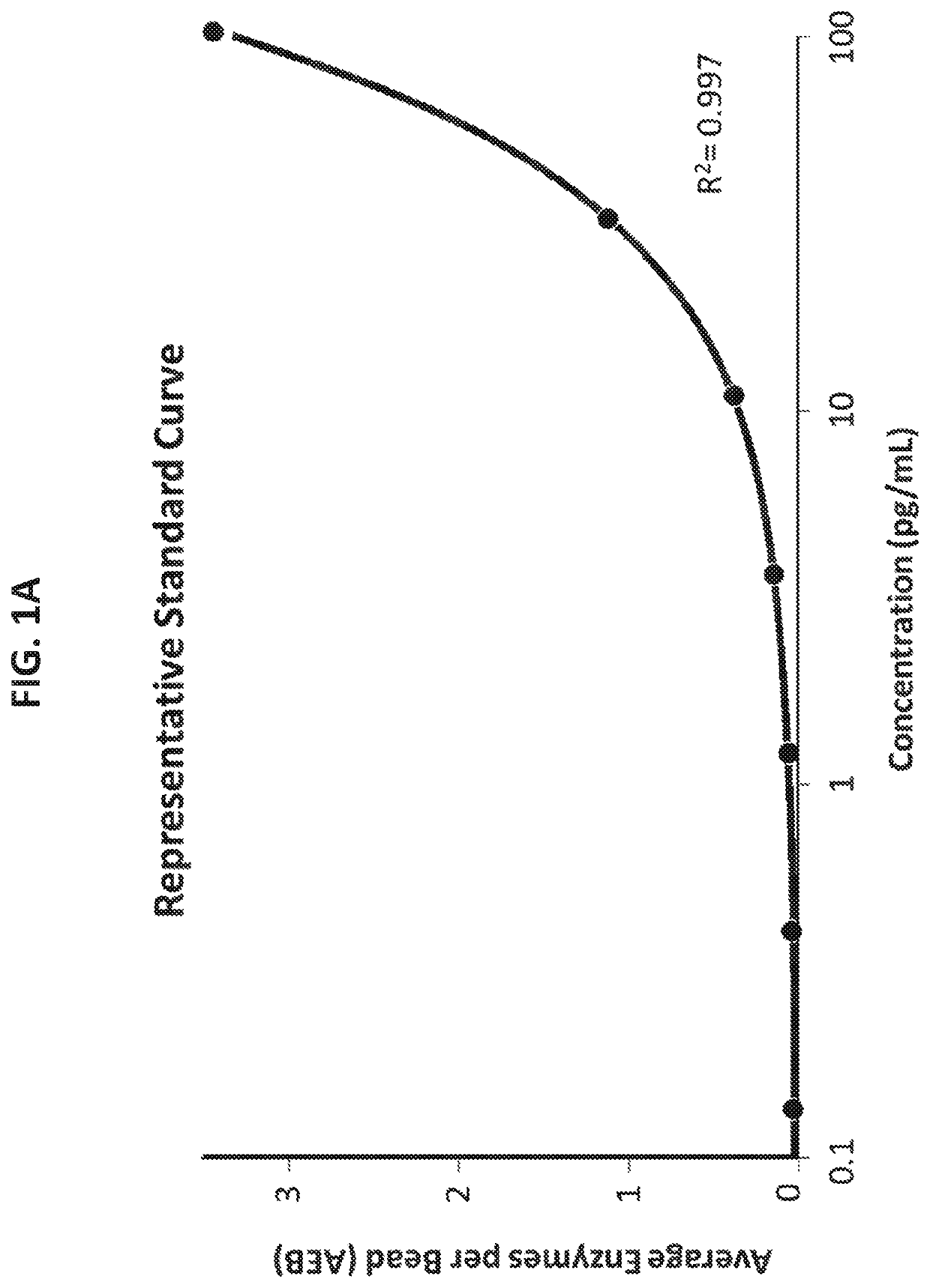
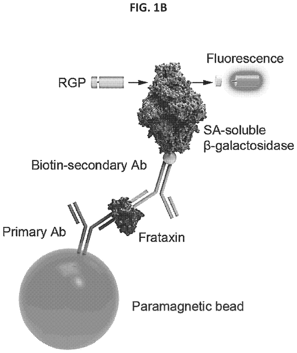
[0409]The single molecule array technology employed two primary steps: an initial analyte capture step conducted with paramagnetic beads, followed by isolation of individual beads in arrays of femtoliter-sized reaction wells for digital imaging. Isolation of the individual beads in microwells permits the buildup of fluorescent product from the enzyme label such that signal from a single immunocomplex is readily detected using a CCD camera, such as the Simoa® HD-1 Analyzer. This approach permits counting of single molecules when frataxin protein concentrations are low enough that the ratio of bound labeled peptide per bead is much less than one. In this concentration realm, Poisson statistics predict that bead-containing microwells in the array will contain either a single labeled frataxin protein molecule or no labeled frataxin protein molecules, resulting in a binary signal. Due to the amplified sensitivity for detecting label molecules afforded by confining ...
PUM
| Property | Measurement | Unit |
|---|---|---|
| Fraction | aaaaa | aaaaa |
| Fraction | aaaaa | aaaaa |
| Fraction | aaaaa | aaaaa |
Abstract
Description
Claims
Application Information