Proteogenomic methods for diagnosing cancer
a proteogenetic method and cancer technology, applied in the field of molecular biology, cell biology, genetics, medicine, can solve the problems of limiting translational research opportunities and the amount of tissue required
- Summary
- Abstract
- Description
- Claims
- Application Information
AI Technical Summary
Benefits of technology
Problems solved by technology
Method used
Image
Examples
example 1
Development and Evaluation of the Biopsy Trifecta Extraction Protocol (Biotext)
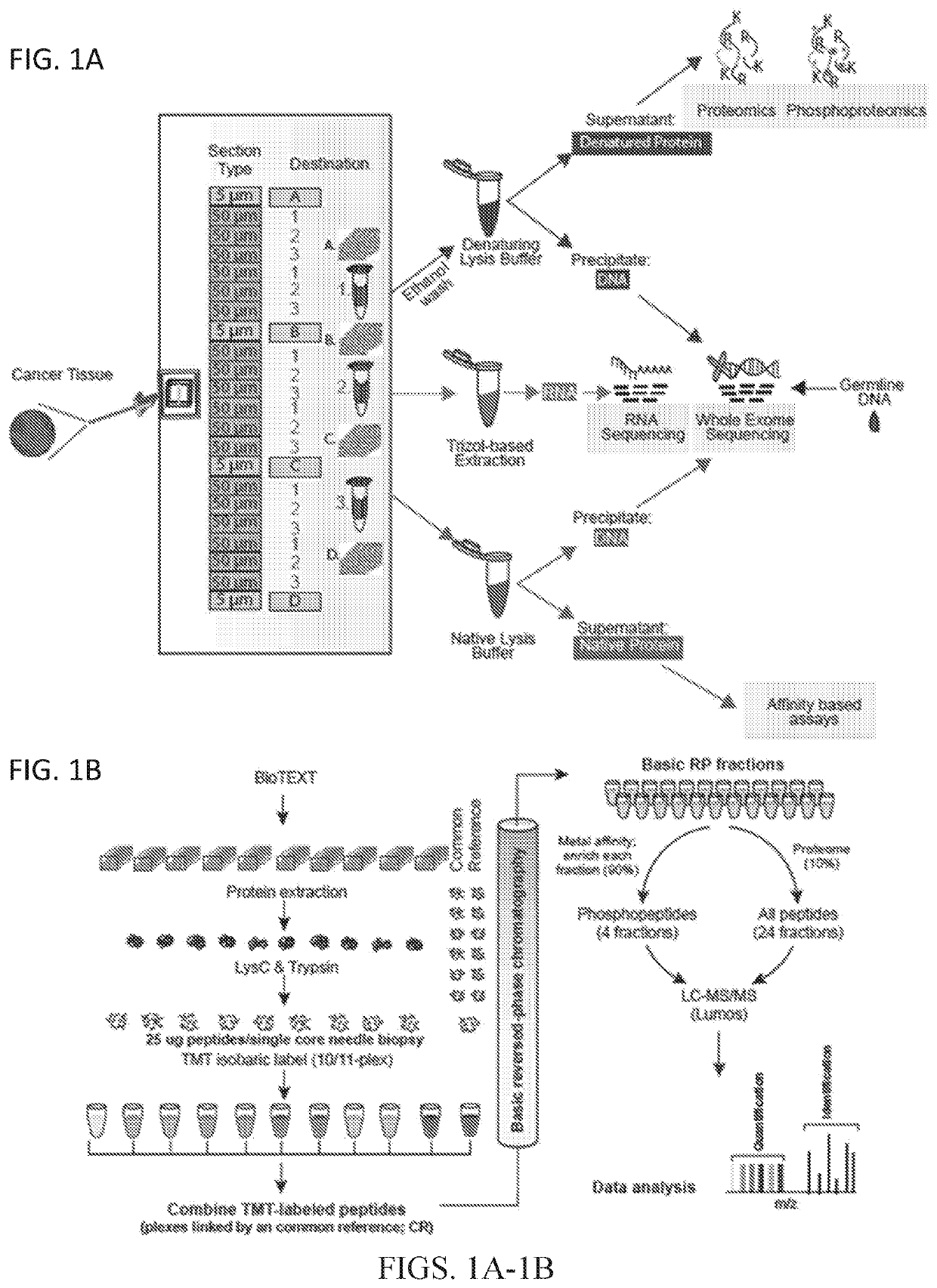
[0085]To perform proteogenomics analyses from flash-frozen diagnostic core needle biopsies, the BioTExt protocol was devised and optimized. A single optimal cutting temperature (OCT)-embedded core biopsy was serially sectioned with alternating 50 μm sections transferred into 3 different 1.5 ml tubes (FIG. 1A). A total of 6 sections were transferred into each tube. To assess sample quality, 5 μm sections were taken before the first and after every sixth 50 μm section for H&E staining, with adequate quality control requiring 50% average tumor content throughout the sample. The first tube was used to extract denatured protein and DNA, the second tube was used for RNA isolation, and the third tube was used to extract native protein and DNA. The denatured protein was subsequently used for proteomic and phosphoproteomic analyses described herein, and DNA and RNA were used for genomic analysis.
example 2
Development and Evaluation of Microscaled Proteomics
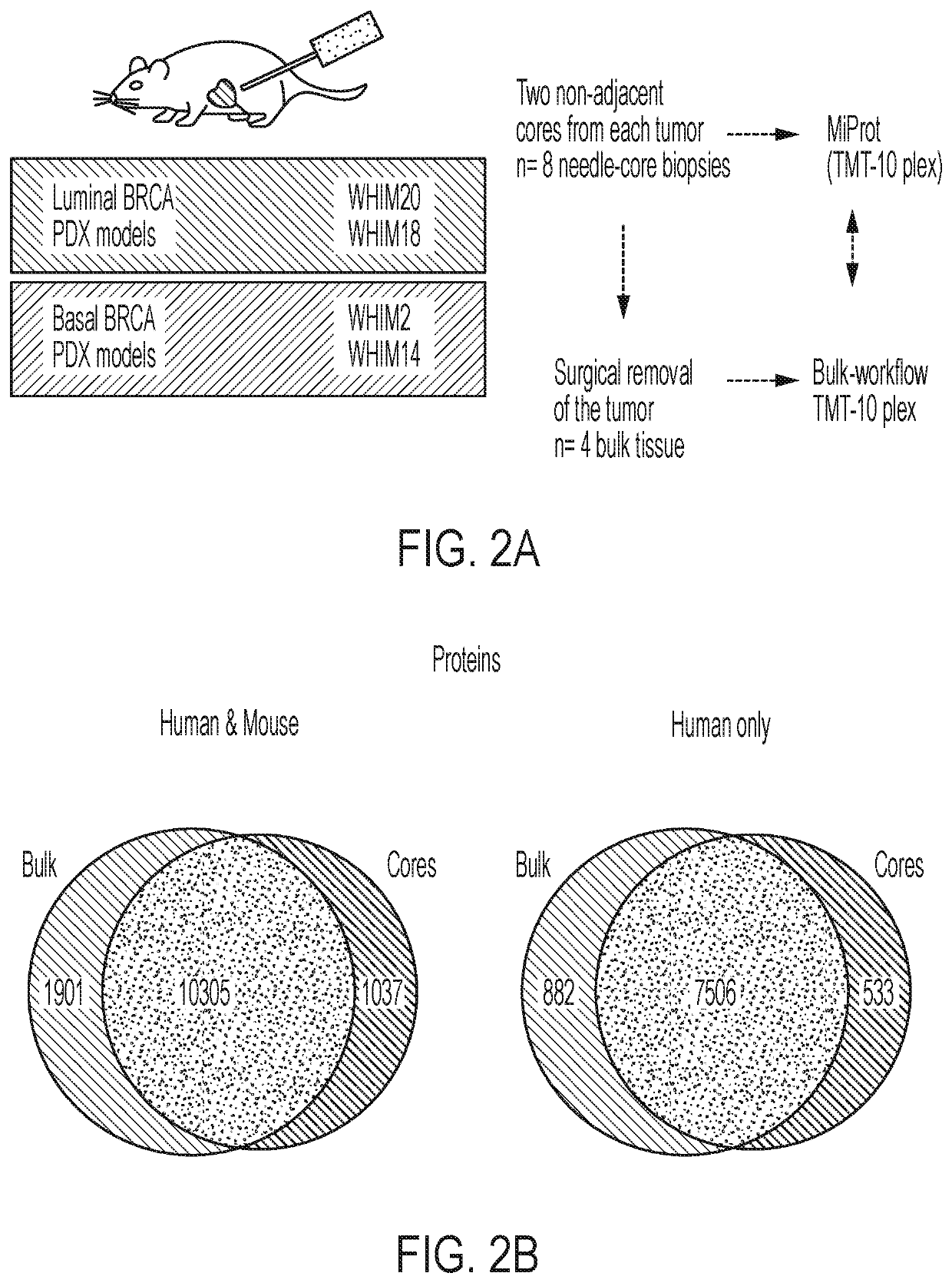
[0086]To assess the quantity of recoverable analytes using the procedure outlined above, BioTExt was applied to several OCT-embedded core-needle biopsies collected from a total of 4 previously established breast cancer patient-derived xenograft (PDX) models: WHIM2, WHIM14, WHIM18 and WHIM2011. The yield for the sum of all six sections from a single biopsy in these PDX tumors ranged from 2.5-14 μg DNA, 0.9-2.3 μg RNA and 280-430 μg of protein. Extraction yields for the nucleic acid extractions are provided in FIG. 6A. The yields of the three analytes required a method capable of providing a deep-scale proteome and phosphoproteome despite lower analyte input. Because a wide range of needle sizes (14-22 gauge) are used to obtain diagnostic biopsies and different tumor types yield widely varying amounts of protein, a minimum of 25 μg of input peptide / sample was set as the target. This amount should reasonably and consistently be obtain...
example 3
Application of Microscaled Proteogenomic Analyses to Clinical Core Biopsies from Patients Treated for ERBB2+ Locally Advanced Breast Cancer
[0091]The effectiveness of the BioTExt and MiProt analyses in PDX models encouraged the application of these methods to clinical tumor samples acquired in the context of a small-scale ERBB2+ breast cancer study (Discovery protocol 1 (DP1); NCT01850628). This study was designed primarily to investigate the feasibility of proteogenomic profiling before and immediately after initiating trastuzumab-based treatment for ERBB2+ breast cancer. Patients with a palpable breast mass diagnosed as ERBB2 positive breast cancer by a local laboratory were treated at the physicians' discretion, typically with trastuzumab in combination with pertuzumab and chemotherapy. The regimens included docetaxel or paclitaxel, the former often combined with carboplatin. The protocol (see Clinical Trial NCT01850628 at the Clinical Trials website of the NIH) was designed to st...
PUM
| Property | Measurement | Unit |
|---|---|---|
| Mass | aaaaa | aaaaa |
| Hydrophobicity | aaaaa | aaaaa |
Abstract
Description
Claims
Application Information
 - R&D
- Intellectual Property
- Life Sciences
- Materials
- Tech Scout
- Unparalleled Data Quality
- Higher Quality Content
- 60% Fewer Hallucinations
Browse by: Latest US Patents, China's latest patents, Technical Efficacy Thesaurus, Application Domain, Technology Topic, Popular Technical Reports.
© 2025 PatSnap. All rights reserved.Legal|Privacy policy|Modern Slavery Act Transparency Statement|Sitemap|About US| Contact US: help@patsnap.com


