Separation and identification of water and fat MR images at mid-field strength with reduced T2/T2* weighting

a technology of mr images and weighting, applied in the field of mr image separation and identification of water and fat at mid-field strength, can solve the problems of requiring at least three mr scans, slow approaches, and inability to completely achieve mr images, and achieve the effect of reducing t.sub.2 and t.sub.2 * weighting

Inactive Publication Date: 2003-08-05
TOSHIBA AMERICA MRI
View PDF11 Cites 32 Cited by
  • Summary
  • Abstract
  • Description
  • Claims
  • Application Information

AI Technical Summary

Benefits of technology

An MRI technique has been developed for distinguishing and uniquely identifying water and fat regions, and for doing so with relatively-low T.sub.2 and T.sub.2 * weighting. This MRI technique breaks the sampling symmetry of prior techniques for distinguishing water and fat signals, such as is described in the '119 patent. The sampling symmetry refers to the acquisition of image data from water and fat signals that are either parallel (in-phase) or anti-parallel (180.degree. out-of-phase) at the centers of all echo signals. To break the sampling symmetry, the present technique acquires two echo images with water and fat signals that are orthogonal (90.degree. out-of-phase) and parallel / anti-parallel, respectively. By breaking the sampling symmetry and acquiring two echo images, the present technique is able to acquire echo signals in a shorter period of time, thus reducing T.sub.2 and T.sub.2 * weighting. By acquiring an echo signal in which water and fat signals are orthogonal, water and fat images can be absolutely identified according to their difference in precessing frequencies, as described later.
c) removing the effect of background field inhomogeneties according to the unwrapped field map to yield a corrected first MR echo signal and a corrected second MR echo signal; d) calculating an intermediate data for the corrected first MR echo image according to the sequence parameters of data acquisition; e) summing the imaginary portion of the intermediate data; f) distinguishing whether a seed reference used in applying the phase unwrapping is water-dominant or fat-dominant based on the polarity of the sum of the imaginary portion of the intermediate data, and g) determining water and fat images from the corrected first and second MR echo images based on whether the seed reference is water-dominant or fat-dominant.

Problems solved by technology

Distinguishing water and fat regions using MR imaging techniques is difficult.
A problem in using the original Dixon method occurs because of inhomogeneities in the static B.sub.O field.
Prior approaches for separating images of water and fat have not been entirely successful in generating MR images that provide good contrast between fat and water regions.
Some approaches are slow and require at least three MR scans.
These approaches with long acquisition times are susceptible to motion problems that arise if the patient moves during the scan period.
Approaches that use single scans (and have fast scan periods) have long minimum echo periods, which result in heavy T.sub.2 / T.sub.2.sup.
* weighting is undesirable when strong T.sub.1 weighting is desired.
Some single scan approaches, such as that disclosed in the '119 patent, do not uniquely identify water and fat nuclei images after separation.
Although MR images of both water and fat may contain the same or different diagnostic information, they often interfere with each other's interpretation when overlapped in an MRI image and thus make it difficult to properly interpret the composite MR image.
However, at mid- or low-field strengths, approaches based on chemical shift selectivity become impractical, if not impossible.
At all-field strengths, the difficulties of water / fat image separation are further exacerbated when there are large magnetic field inhomogeneities.
The difficulty in separating fat and water images in a practical MR imaging application is particularly true for mid- and low-field systems where the frequency separation between the water and fat signals is much reduced in comparison to that at high fields.
Such long scan periods are susceptible to motion problems.
This 180.degree. angular difference makes it difficult to uniquely identify fat and water images after separation, for reasons described below.
Moreover, there is no other information available to automatically distinguish the water and fat signals.

Method used


Image

  • Separation and identification of water and fat MR images at mid-field strength with reduced T2/T2* weighting
  • Separation and identification of water and fat MR images at mid-field strength with reduced T2/T2* weighting
  • Separation and identification of water and fat MR images at mid-field strength with reduced T2/T2* weighting

Examples


case 1 (

when the reference is water-dominant): ##EQU9##

case 2 (

when the reference is fat-dominant): ##EQU10##

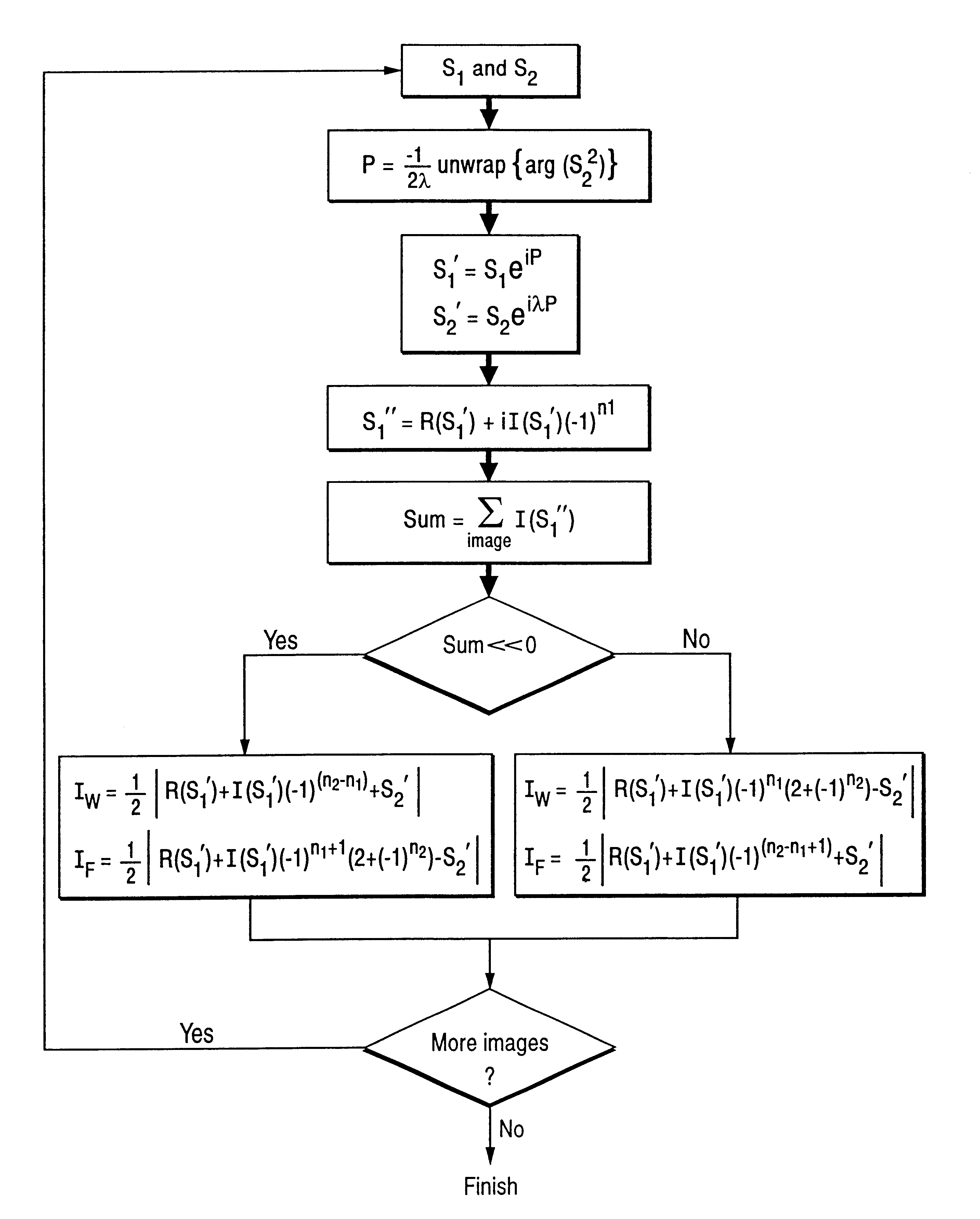
A complete data processing procedure for separating and identifying water and fat images is summarized schematically in the flowchart shown in FIG. 7.

One implementation of the above-described method of data acquisition is a field-echo sequence which acquires two echo images with n.sub.1 =0 and n.sub.2 =1, such that .lambda.=2. At 0.35 Tesla, then TE.sub.1 =4.75 ms and TE.sub.2 =9.50 ms for water and fat separation. The sequence can be used in either two-dimensional (2D) or three-dimensional (3D) imaging modes. FIGS. 8 and 9 are diagrams of the pulse sequencer for such 2D and 3D data acquisition.

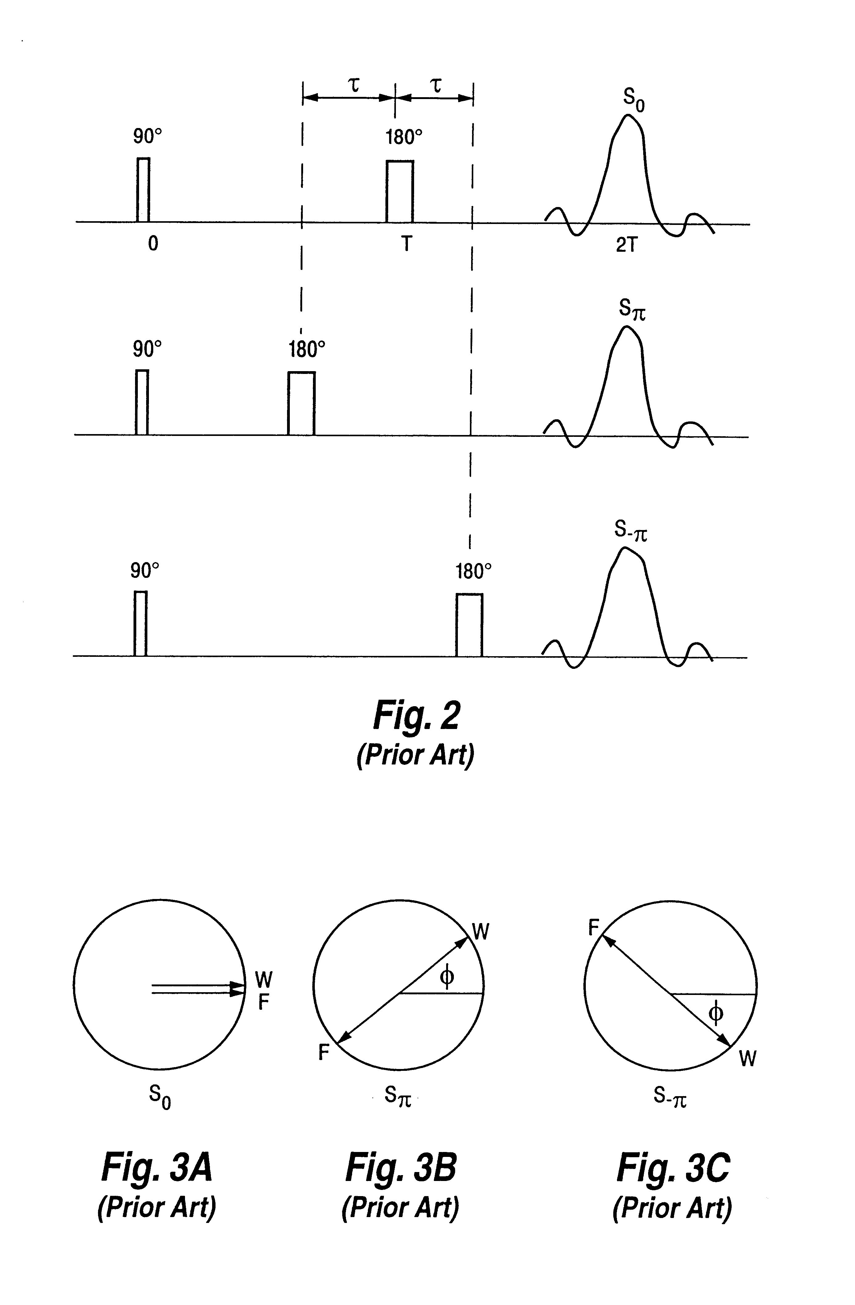
Referring to FIG. 6, a spin-echo imaging sequence of the present invention basically consists of an RF nuclei nutation ("excitation") pulse, 40, accompanied by the generation of a slice selection gradient pulse, G.sub.slice, and a phase encoding gradient pulse, G.sub.pe, followed by three appropriately timed read-out gradient pulses, G.sub.read, 4...


PUM


Abstract

A magnetic resonance imaging (MRI) method is disclosed for generating and identifying water and fat separated MR images. Image data is first acquired to obtain two echo images with the water and fat signals orthogonal in the first echo image, and parallel / anti-parallel in the second echo image. The effect of background field inhomogeneties are removed, and water and fat images are separated from each other. The separated water and fat images are identified according to the difference of their precessing frequencies.

Description

This invention relates generally to magnetic resonance (MR) imaging (MRI) techniques. In particular, it relates to methods for generating water / fat separated images and, more particularly, to a method for generating and identifying water / fat component MR images with reduced T.sub.2 / T.sub.2.sup.* weighting.In broad summary, the present invention relates to MRI techniques for generating images in which fat and water regions can be readily discerned. MRI is often used to non-invasively generate images of the internal organs and other body parts of human patients. It is desirable to distinguish regions of water and fat in an MR image of a patient. Distinguishing water and fat regions using MR imaging techniques is difficult. At mid-field strength the Three-Point Dixon method is the method of choice for separating water and fat images. The original Dixon method for separation of water and fat images depends upon the ability to accurately compensate for inhomogeneities in a polarizing st...

Claims


Application Information

Patent Timeline
Patent Type & Authority Patents(United States)
IPC IPC(8): A61B5/055G01R33/54G01R33/48
CPCA61B5/055G01R33/4828
Inventor ZHANG, WEIGUOKRAMER, DAVID M.
Owner TOSHIBA AMERICA MRI
Why Eureka
Try Eureka