Modified laryngoscope blade to reduce dental injuries during intubation

a laryngoscope and blade technology, applied in the field of modified laryngoscope blades, can solve the problems of inability to completely remove the possibility of intubation trauma, dental damage is a potential, and the maxillary incisors are at particular significant risk during intubation, so as to reduce the likelihood of trauma

Inactive Publication Date: 2003-09-23
CARTLEDGE MEDICAL PRODS
View PDF128 Cites 24 Cited by
  • Summary
  • Abstract
  • Description
  • Claims
  • Application Information

AI Technical Summary

Benefits of technology

As discussed above, there remains a need to further reduce the likelihood of trauma to which all of the mouth and throat are exposed during the technique of tracheal intubation. Therefore, one object of the present invention is to provide a device to allow intubation while adequately protecting the teeth, tongue, and oral mucosa from the blade of a laryngoscope during orotracheal or direct nasotracheal intubation.
Still another object of the present invention is to provide a method for carrying out the technique of intubation whereby the teeth, tongue, and oral mucosa are protected from iatrogenic trauma.
Still another object of the present invention is to provide a device that will permit rapid and easy protection of the teeth, tongue, and oral mucosa from the blade of a laryngoscope during intubation, while requiring minimal additional actions on the part of the user / operator.

Problems solved by technology

Use of a gentle technique for inserting the laryngoscope, may lessen, but cannot totally remove the possibility of intubation trauma.
However, poor dentition or suboptimal anatomy of the teeth, jaw, neck, or throat may leave the maxillary incisors at particularly significant risk during intubation by even the most experienced of technicians.
Because the laryngoscope blade is necessarily formed of a hard, inflexible material, dental damage is a potential result of such intubation procedures.
Even edentulous patients may be at risk for significant injuries to their maxillary alveolar ridges during the process of endotracheal intubation.
The same mechanism of injury that may fracture or dislocate teeth may contuse or fracture the maxillary alveolar ridge, resulting in post-intubation difficulties in using the patient's previous dental prostheses or dentures.
Such prostheses may have to be replaced, at considerable cost.
In addition, residual local pain at the site of injury may aggravate the patient's tolerance for the use of any dental prostheses following such an intubation injury.
Protected shields have limited advantage in preventing dental injuries during intubation.
The shields cover the maxillary incisors, yet provide only limited protection for the teeth.
Such shields are prone to displacement by either the laryngoscope blade or other instrumentation.
Furthermore, existing shields are relatively difficult to use, requiring multiple operator steps to secure the shields for use.
The multiple steps required to secure existing shields may discourage their use by significantly increasing the time and effort required to achieve the desired intubation.
Moreover, the bulk of the protective shields may obstruct the user's view into the hypopharynx.
The devices currently available do not satisfactorily address the need to protect a patient's teeth, as well as the needs of convenience and feasibility of use.
Blade covers are cumbersome and slow to apply to the blade, and are therefore impractical in emergency situations.
Adhesives used in applying a blade cover to a blade often make it difficult to remove the blade cover and may leave a residue that is difficult to remove.
Adhesive residues may become even more difficult to remove after the residue has been subjected to the heat and pressure of the sterilization process.
In addition, any such residue on a reusable instrument may present a potential focus for infectious bacteria or other pathogenic organisms.
Further, blade covers that surround the entire blade or entire flange occupy too much space in a patient's mouth, making manipulation of the laryngoscope blade and introduction of the endotracheal tube difficult.
Common user errors during positioning of a laryngoscope, as well as the particular structure of a given blade, may cause difficulties in proper introduction of an endotracheal tube.
Such errors, which include the placement of the blade at an improper depth and incomplete displacement of the tongue, may be more likely with one blade over another.
For example, complete displacement of the tongue may be more difficult with blades styles wherein the flange curves back over the blade, presenting a relatively small passageway for the actual passage of an endotracheal tube.

Method used


Image

  • Modified laryngoscope blade to reduce dental injuries during intubation
  • Modified laryngoscope blade to reduce dental injuries during intubation
  • Modified laryngoscope blade to reduce dental injuries during intubation

Examples


Embodiment Construction

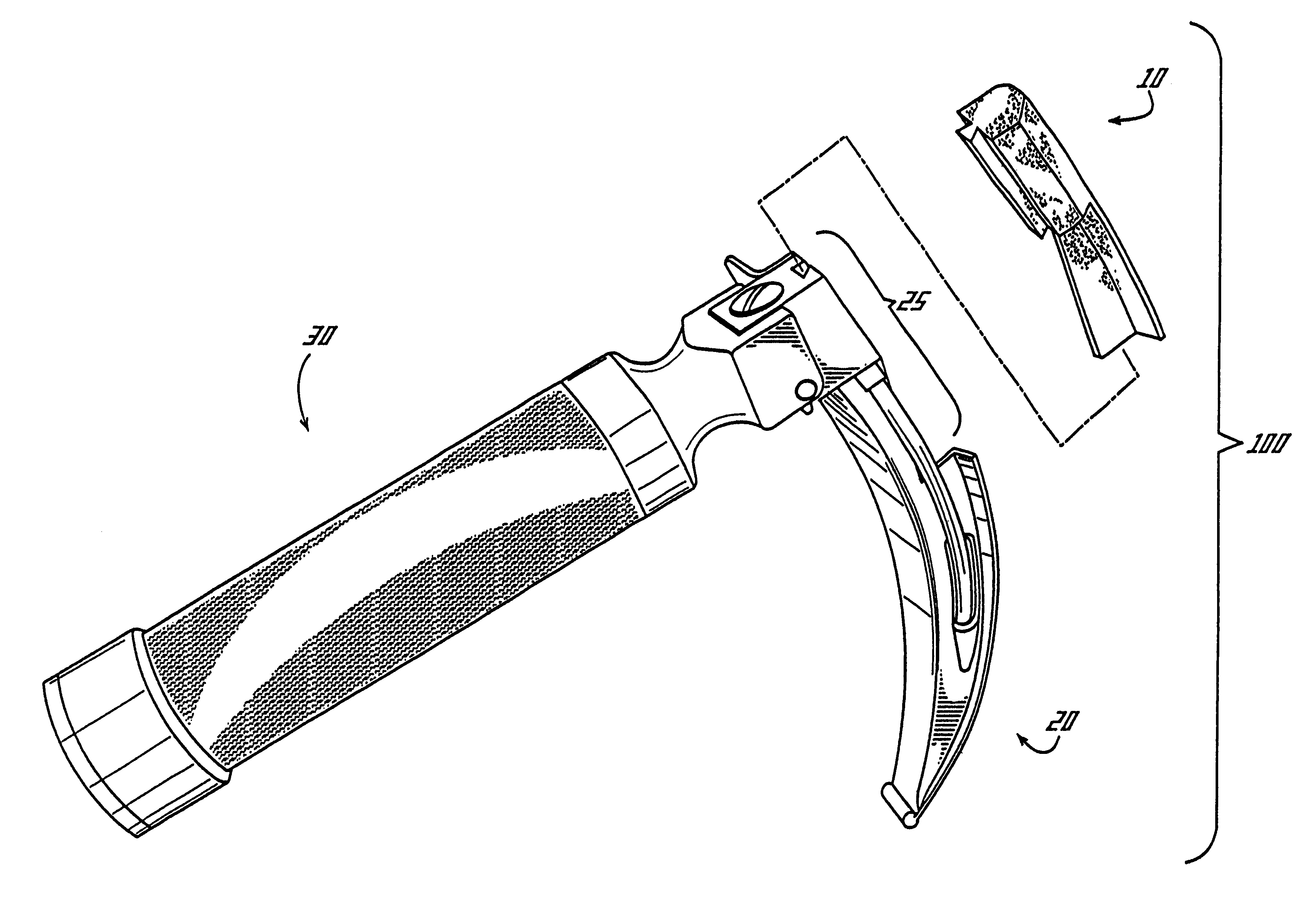
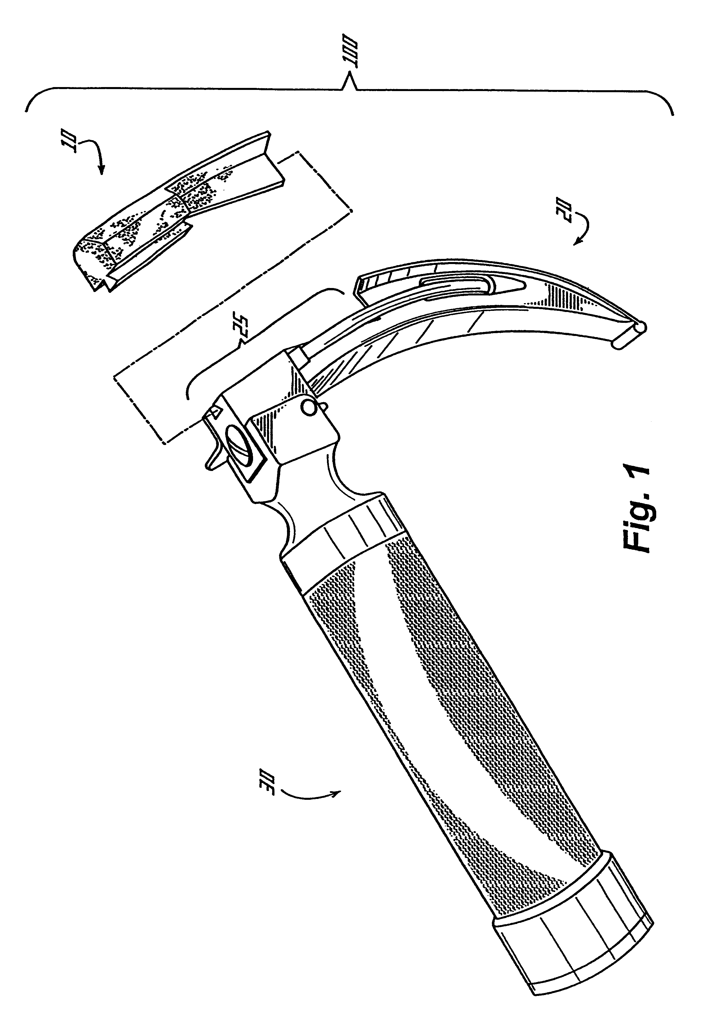
An overview of a preferred embodiment of the present invention is illustrated in FIG. 1. The inventive modified laryngoscope blade assembly 100 includes a removable protective insert 10 and a modified blade structure 20. The blade structure 20 has a recess 25 in its upper surface into which the insert 10 is received. The inventive modified laryngoscope blade assembly 100 is designed to functionally mount on a conventional laryngoscope handle 30 which serves both as a user handle and as a battery housing and power supply.

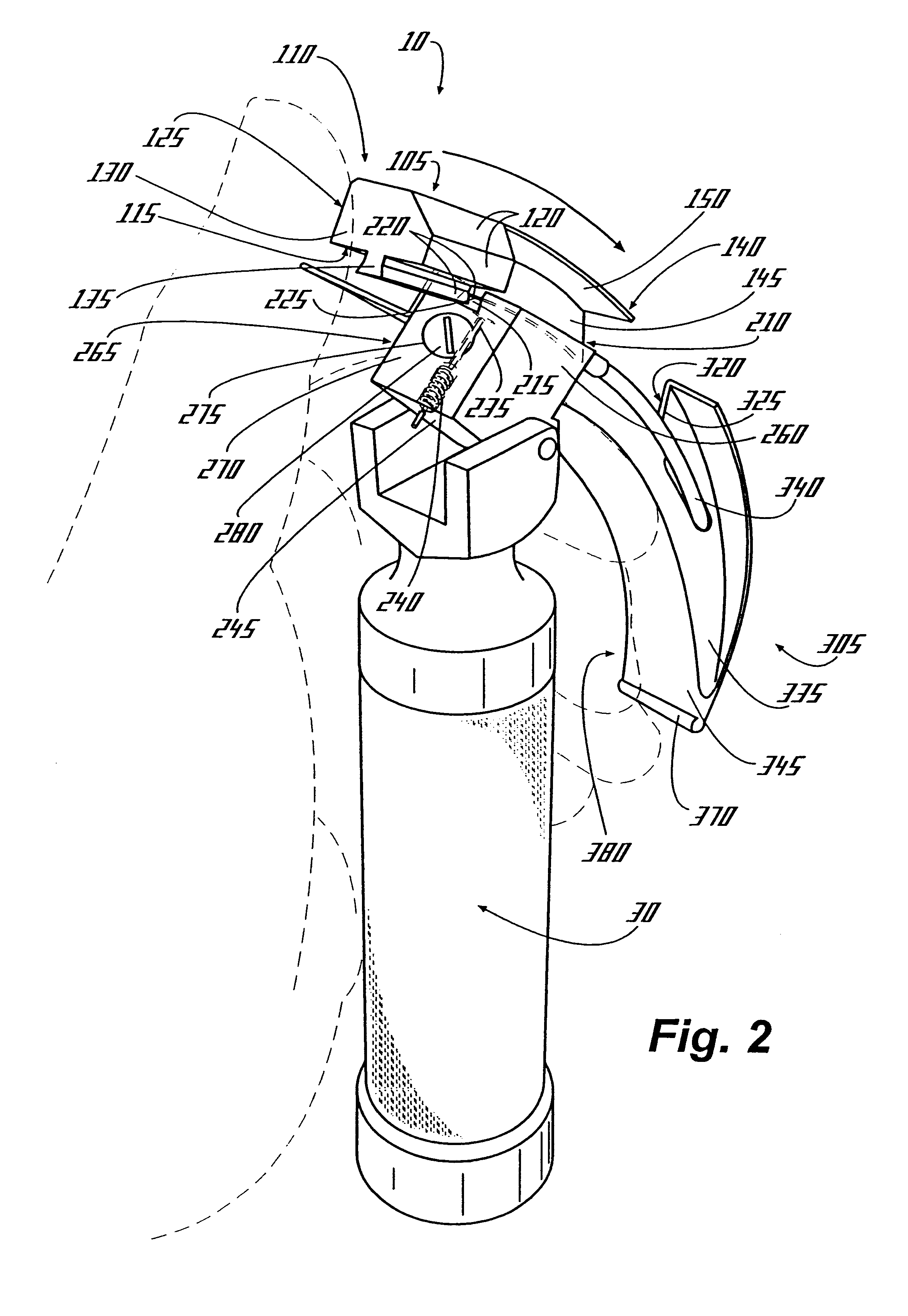
FIG. 2 provides further details of the working interaction among the removable protective insert 10, the modified blade structure 20, and the conventional laryngoscope handle 30 of FIG. 1. The protective insert 10 has a body portion 105 and a blade portion 145. The body portion 105 has an maxillary surface 110, a blade interface surface 115, a buccal surface 120, a medial surface 125, an anterior surface 130, and a retention ridge 135. The retention ridge 135 extends...


PUM


Abstract

The present invention relates to the field of medical devices used in the procedures of orotracheal or nasotracheal intubation. Oral or nasal endotracheal intubation procedures are commonly employed to secure a controlled airway and to deliver inhalant oxygen, anesthetic gases, and other therapeutic agents into the trachea and lungs of human and veterinary patients. Such intubation procedures carry a significant risk of dental injury resulting from contact between the laryngoscope blade used for visualization during intubation. The present invention provides an apparatus to reduce dental injury including a modified laryngoscope blade and a disposable insert which is designed to be received and retained in a single step by the modified laryngoscope blade. The disposable insert may be quickly secured by the user, and reduces both direct pressure and shear forces on the maxillary incisor teeth when the laryngoscope blade is placed in a patient's mouth during intubation.

Description

The present invention relates to the field of medical devices used in the procedures of orotracheal or nasotracheal intubation. More particularly, the present invention relates to a modified laryngoscope blade which is designed to receive a disposable insert which reduces both direct pressure and shear forces on the maxillary incisor teeth when the laryngoscope blade is placed in a patient's mouth during intubation.Oral or nasal endotracheal intubation procedures are commonly employed to secure a controlled airway and to deliver inhalant oxygen, anesthetic gases, and other therapeutic agents into the trachea and lungs of human and veterinary patients. These clinical procedures are usually performed as a preliminary measure before surgery, thus enabling the anesthesiologist to administer anesthetic gases and other drugs while maintaining a stable delivery of oxygen to the patient's lungs throughout the surgical procedure. In other settings, intubation is used to provide a patient wit...

Claims


Application Information

Patent Timeline
Patent Type & Authority Patents(United States)
IPC IPC(8): A61B1/267
CPCA61B1/267
Inventor CARTLEDGE, RICHARDLANE, JOSHUA
Owner CARTLEDGE MEDICAL PRODS
Why Eureka
Try Eureka