Implantable article and method for treating urinary incontinence using means for repositioning the implantable article

a technology of implantable articles and urinary incontinence, which is applied in the field of implantable articles and methods for treating urinary incontinence, can solve the problems of incontinence type, functional incontinence, and difficulty in moving from one place to another, so as to avoid permanent deformation of the sling, and adversely affect the therapeutic effect of the sling

Inactive Publication Date: 2003-11-25
STASKIN DAVID MD DR
View PDF154 Cites 629 Cited by
  • Summary
  • Abstract
  • Description
  • Claims
  • Application Information

AI Technical Summary

Benefits of technology

The present invention comprises an implantable article suitable for treating urological disorders in patients. In one aspect, the present invention comprises a sling for treating urinary incontinence in a patient. The sling has first and second major surfaces, a pair of end portions, and a support portion for placement in a therapeutically effective position relative to a physiological environment intended to be supported. The support portion preferably has an axially elongate mesh, and a pair of ends. The present invention includes repositioning means, associated with the sling, for transferring either sling tightening or sling loosening forces along the sling to afford effective, permanent repositioning of the sling without adversely affecting the therapeutic effect of the sling. The repositioning means transfers sling tightening or sling loosening forces along the sling while avoiding permanent deformation of the sling. Preferably, the repositioning means is constructed to afford transfer at least some of a repositioning force applied to the sling to an end of the support portion.
The elements of the assembly of the present invention may be any color. Preferably, the elements are of constructed to be a color that contrasts with the intended physiological environment and with other elements. For example, the sling 42 is preferably white and the position adjustment member 66 may be blue. This helps the surgeon identify the location and discern the elements of the assembly.

Problems solved by technology

Other factors, such as trauma to the urethral area, neurological injury, hormonal imbalance or medication side-effects, may also cause or contribute to incontinence.
Inappropriate bladder contractions and weakened sphincter muscles usually cause this type of incontinence.
Functional incontinence results when a person has difficulty moving from one place to another.
Although serious complications associated with sling procedures are infrequent, they do occur.
Complications associated with the TVT procedure and other known sling procedures include injury to blood vessels of the pelvic sidewall and abdominal wall, hematomas, urinary retention, and bladder and bowel injury due to passage of large needles.
One serious disadvantage of the TVT procedure, particularly for surgeons unfamiliar with the surgical method, is the lack of information concerning the precise location of the needle tip relative to adjacent pelvic anatomy.
If the needle tip is allowed to accidentally pass across the surface of any blood vessel, lymphatic duct, nerve, nerve bundle or organ, serious complications can arise.
Additional problems are associated with the TVT and other sling procedures.
However, the smooth surface of the needles, which facilitates insertion through the tissues, prevents secure attachment of the forceps onto the needles, causing slippage or detachment of the forceps during the withdrawal procedure.
Removal and reuse of the handle of the TVT product is also a cumbersome, time consuming process, requiring the surgeon to manually rotate the handle until the handle is unscrewed from the needle.
Reusing the handle presents a contamination risk, particularly if the handle and screw threads are not properly cleaned and sterilized after use on one side of the patient.
The problems associated with improper placement of the TVT mesh are particularly troublesome.
If the mesh is too loosely associated with its intended physiological environment, the mesh may be ineffective in supporting the urethra and treating incontinence.
Several complications can arise from a mesh that is too tightly placed including retention, sling erosion and other damage to surrounding tissue such as the urethra and vagina.
Once the sheath is removed from the mesh of the TVT product, friction between the mesh and tissue keeps the mesh in position and it becomes very difficult to subsequently adjust the position of the mesh relative to tissue.
If insufficient adjustment force is applied, the sling will simply exhibit a memory property and return to its original, unacceptable position.
As a result, surgeons are tempted to use a great deal of force in order to loosen a sling that is perceived to be too tightly associated with its intended physiological environment.
Under excessive deformation, the holes of the TVT mesh become significantly smaller, and risk deterring tissue ingrowth.
In the excessively deformed state, the edges of the mesh tend to curl up and present a relatively sharp, frayed surface.
In this curled or deformed state, the edges of the TVT mesh present sharp surfaces that can readily abrade or otherwise damage adjacent tissue such as the urethra, bladder or vagina.
Attempts to reposition the TVT sling is likely to fail in two modes.
First, the surgeon may apply insufficient elongation force to the mesh (e.g. with forceps), resulting in temporary elastic deformation of the mesh followed by a return by the mesh to its original, unacceptable position after the force is removed.
Second, the surgeon may apply excessive force to the mesh resulting in the curling deformation described above with the attendant risk of tissue damage.
Thus, even if the edges do not curl, excessive deformation of the TVT sling risks adversely affecting sling performance.

Method used


Image

  • Implantable article and method for treating urinary incontinence using means for repositioning the implantable article
  • Implantable article and method for treating urinary incontinence using means for repositioning the implantable article
  • Implantable article and method for treating urinary incontinence using means for repositioning the implantable article

Examples


Embodiment Construction

OF METHODS

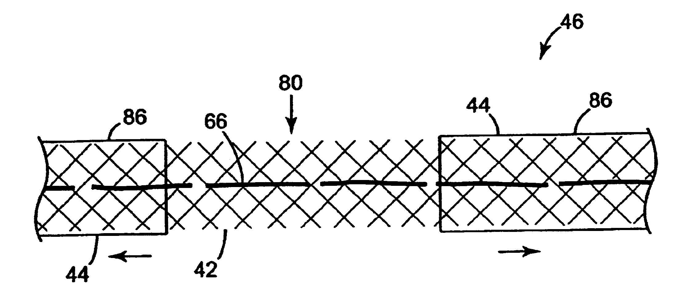
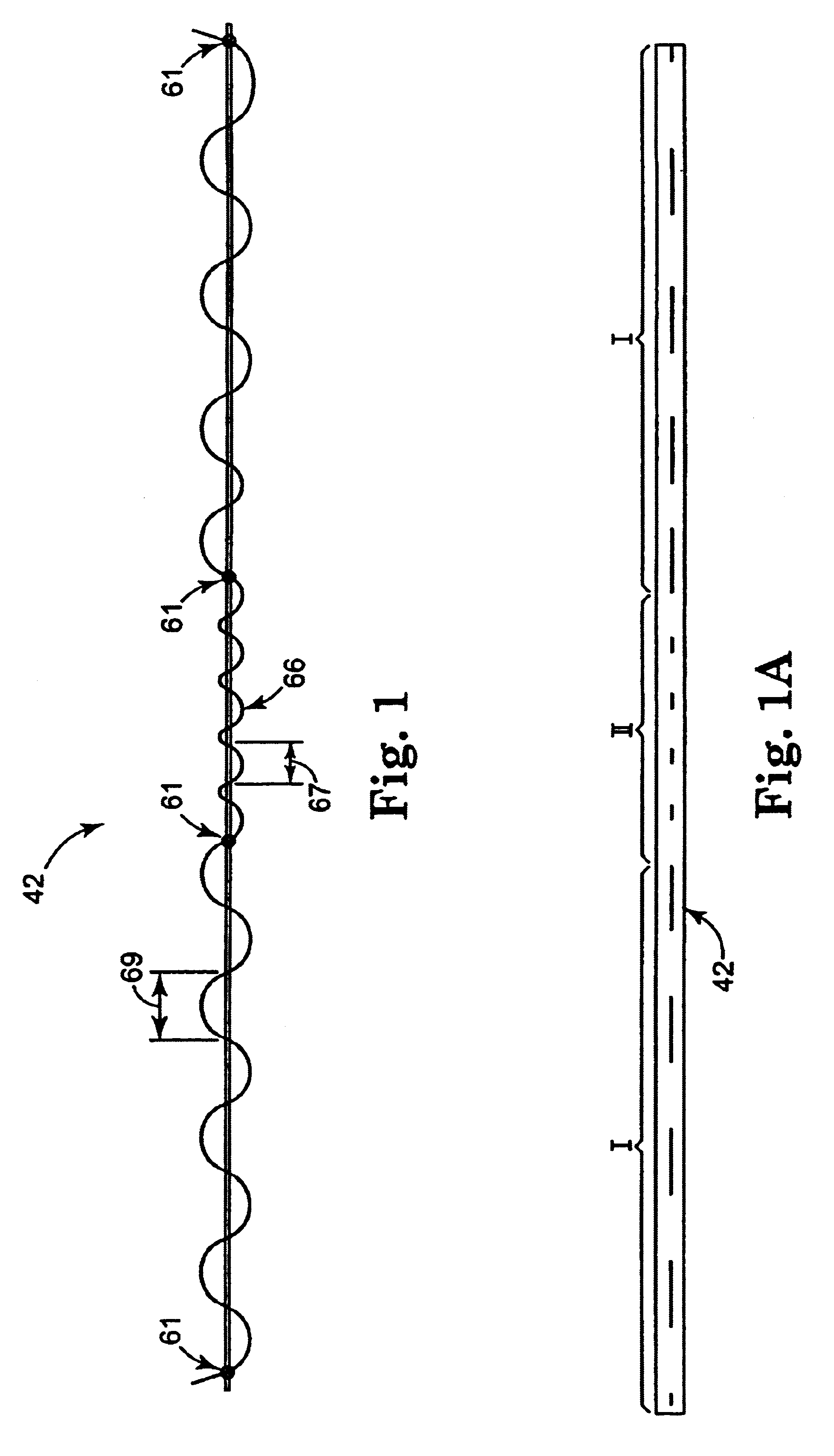
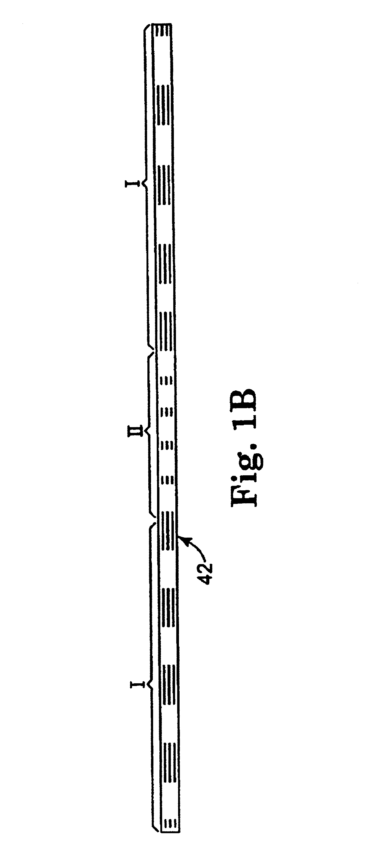
Many methods are contemplated herein. Although the methods of use as disclosed herein generally relate to female incontinence conditions and treatments / procedures, male incontinence conditions and treatments / procedures are also included within the scope of the present invention. Procedures that address problems other than incontinence (e.g. cystocele, enterocele or prolapse) are also contemplated alone or in conjunction with the present invention. Further, the term "urethra," with respect to sling positioning, is used for brevity and reader convenience. It should be noted that the present invention is particularly suitable for placing a sling in a therapeutically effective position. The method may be utilized to support a variety of structures at different anatomical locations. As such, the terms "target site," "bladder", "urethro-vesical juncture", "vaginal vault", "U-V juncture" and "bladder neck" are also included within the scope of the present invention.

Referring now ...


PUM


Abstract

An implantable article and method of use are disclosed to treat urological disorders. The biocompatible device includes a sling assembly configured to be minimally invasive and provide sufficient support to the target site. In addition, the configuration of the sling assembly also allows the position of the sling to be permanently changed during and / or after implantation.

Description

Over 13 million American men and women of all ages suffer from urinary incontinence. The social implications for an incontinent patient include loss of self-esteem, embarrassment, restriction of social and sexual activities, isolation, depression and, in some instances, dependence on caregivers. Incontinence is the most common reason for institutionalization of the elderly.The urinary system consists of the kidneys, ureters, bladder and urethra. The bladder is a hollow, muscular, balloon-shaped sac that serves as a storage container for urine. The bladder is located behind the pubic bone and is protected by the pelvis. Ligaments hold the bladder in place and connect it to the pelvis and other tissue. FIG. 2 schematically illustrates female anatomy. The urethra 16 is the tube that passes urine from the bladder 14 out of the body. The narrow, internal opening of the urethra 16 within the bladder 14 is the bladder neck 18. In this region, the bladder's bundled muscular fibers transitio...

Claims


Application Information

Patent Timeline
Patent Type & Authority Patents(United States)
IPC IPC(8): A61F2/00A61B17/06A61B17/04A61B17/32A61B17/30A61B17/42A61B19/02A61B19/00A61B17/00A61F2/02A61F13/00
CPCA61F2/0045A61B17/0482A61B17/0487A61B17/06004A61B17/06109A61B17/0401A61B17/04A61B17/0469A61B17/06066A61B17/30A61B17/3211A61B17/42A61B19/026A61B19/24A61B2017/0046A61B2017/00805A61B2017/06009A61B2017/06014A61B2017/06042A61B2017/06085A61B90/02A61B50/30
Inventor NEISZ, JOHANN J.WESTRUM, JR., JOHN W.PORTER, CHRISTOPHER H.
Owner STASKIN DAVID MD DR
Why Eureka
Try Eureka