Method of unsupervised cell nuclei segmentation

a cell nucleus and cell nucleus technology, applied in the field of unsupervised cell nucleus segmentation, can solve the problems of difficult to achieve high degree of accuracy in very large data sets, false positive and false negative indications cannot be tolerated, and the stain used in the papanicolaou slide preparation process does not produce a strong nucleus-cytoplasm contrast. , to achieve the effect of improving the method of image segmentation

Inactive Publication Date: 2004-01-20
COOP RES CENT FOR SENSOR SIGNAL & INFORMATION PROCESSING IN THIS DEED COLLECTIVELY CALLED CSSIP
View PDF7 Cites 21 Cited by
  • Summary
  • Abstract
  • Description
  • Claims
  • Application Information

AI Technical Summary

Benefits of technology

It is an object of the invention to provid

Problems solved by technology

Obviously, false positive and false negative indications cannot be tolerated.
However, a high degree of accuracy in very large data sets has proven to be difficult to achieve.
Unfortunately, the stain used in the Papanicolaou slide preparation process does not produce a strong nucleus-cytoplasm contrast.
Weak image gradients along the nuclear border and artefacts in the cytoplasm add to the difficulty of segmentation.
Even so, an error rate of 1.7% will be unacceptable in many cases.
Previous st

Method used


Image

  • Method of unsupervised cell nuclei segmentation
  • Method of unsupervised cell nuclei segmentation
  • Method of unsupervised cell nuclei segmentation

Examples


Embodiment Construction

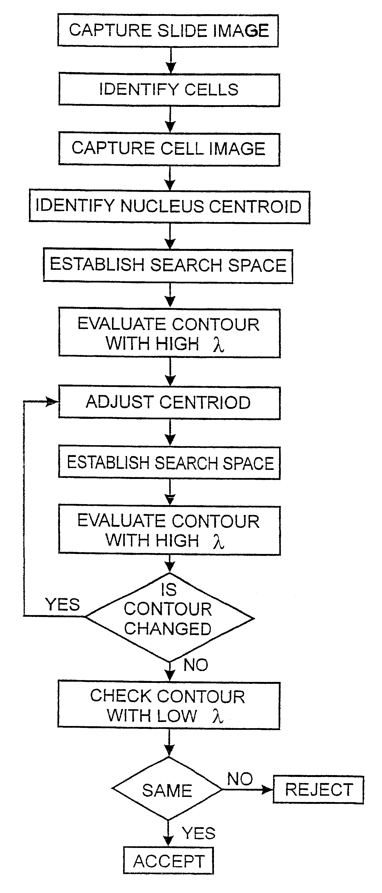
Referring to FIG. 1 there is shown a flow chart of the process for cell nuclei segmentation. The method is described with reference to the preferred embodiment of identifying and segmenting cell nuclei in a pap smear slide. It will be appreciated that the steps may be generalised to apply to any segmentation task where very high confidence levels are required.

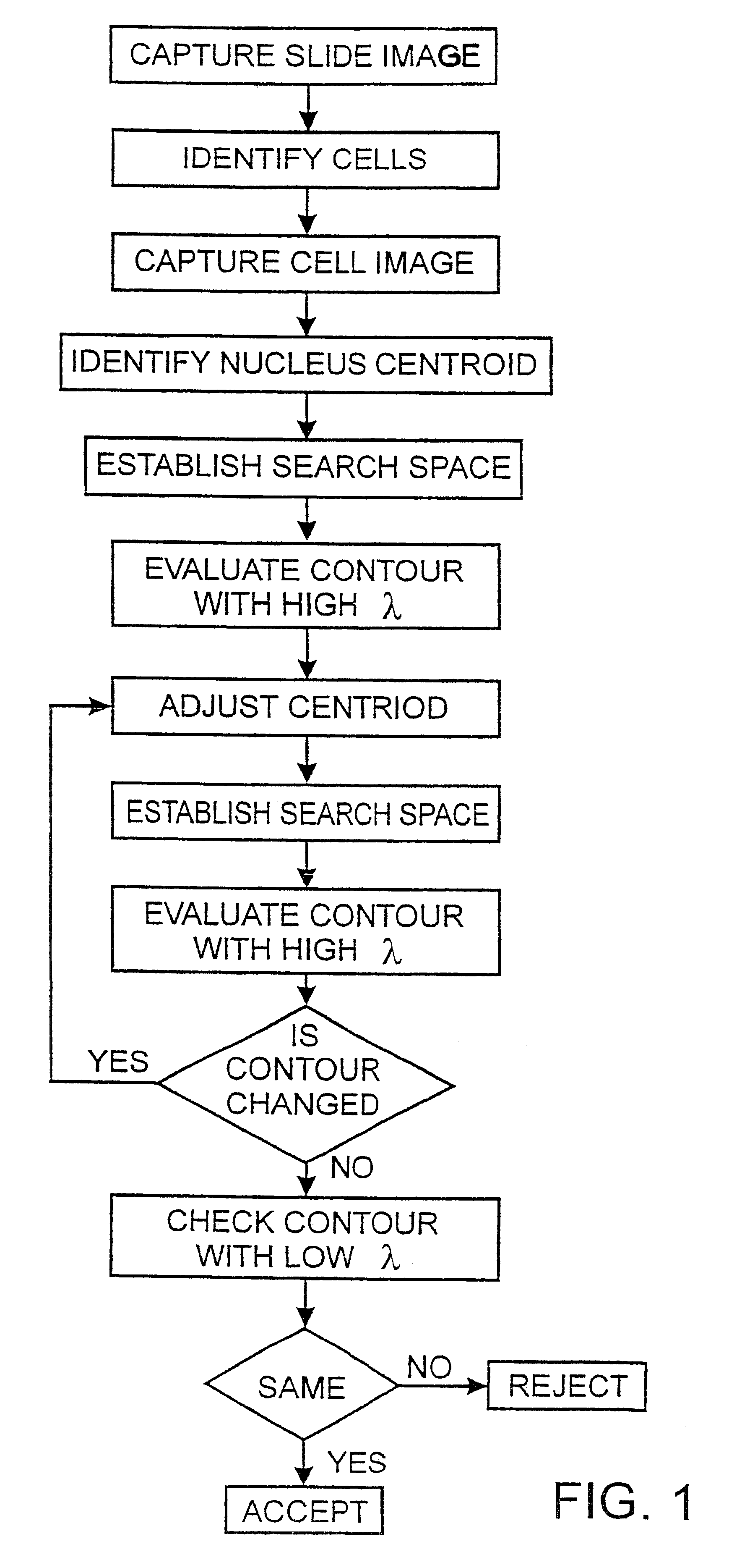
Scenes from a cervical smear slide are captured at low resolution using a CCD camera mounted upon a microscope. Locations of the cells within the image are identified using any suitable technique. One technique is to use the `water immersion algorithm` described by Bamford and Lovell [A water immersion algoritm for cytological image segmentation; APRS Image Segmentation Workshop, University of Technology Sydney, Sydney, December 1996, 75-79]. An example of a scene segmented using the water algorithm is shown in FIG. 2.

Once the cells have been identified a high resolution image is taken of each cell for further processing. The i...


PUM


Abstract

A method of segmentation of cell nuclei is described that uses an active contours approach based on a Viterbi search algorithm. Also disclosed is an initialisation technique that maximises the probability of a correct segmentation. The invention also includes a method to classify cell nuclei according to the difficulty of correct segmentation. Nuclei that are difficult to segment can be rejected to minimise the probability of false positives. The probability of false positives can be driven close to 0%.

Description

This invention relates to an improved method of segmenting objects in an image. In particular, it relates to a method of active contours using a Viterbi search algorithm for unsupervised segmentation of cell nuclei.BACKGROUND TO THE INVENTIONVarious techniques exist for identifying and segmenting objects in an image. Typically an accuracy rate of greater than 90% is considered to be good. However, in biological systems, particularly screening situations, an accuracy rate approaching 100% is required.One application in which a high level of screening confidence is required is the screening of pap smears to identify cervical cancer. Obviously, false positive and false negative indications cannot be tolerated. However, a high degree of accuracy in very large data sets has proven to be difficult to achieve. It has been established by Bengtsson [The measuring of cell features; Anal Quant Cytol; 9 (3) June 1987, 212-217] and others, that segmentation of cell nuclei is the key step in devi...

Claims


Application Information

Patent Timeline
IPC IPC(8): G06T5/00G01N33/48G06F19/00G06K9/00G06K9/64G06T1/00G06T7/00G06T7/12G06T7/143G06T7/149G06T7/60
CPCG06K9/0014G06K9/6207G06T7/0012G06T7/0083G06T7/0087G06T7/0089G06T2207/10056G06T2207/30012G06T2207/30024G06T7/12G06T7/143G06T7/149G06V20/695G06V10/755
Inventor BAMFORD, PASCALLOVELL, BRIAN
Owner COOP RES CENT FOR SENSOR SIGNAL & INFORMATION PROCESSING IN THIS DEED COLLECTIVELY CALLED CSSIP
Why Eureka
Try Eureka