Eureka AIR delivers breakthrough ideas for toughest innovation challenges, trusted by R&D personnel around the world.

Intraosteal ultrasound during surgical implantation

a technology of intraosteal ultrasound and surgical implantation, which is applied in the direction of osteosynthesis devices, catheters, applications, etc., can solve the problems of increasing the risk of breach, increasing the complexity of the task, and injuring critical structures in close proximity, so as to facilitate “real-time” manipulation and navigation of the devi

Inactive Publication Date: 2005-02-01
CUTTING EDGE SURGICAL
View PDF15 Cites 228 Cited by
  • Summary
  • Abstract
  • Description
  • Claims
  • Application Information

AI Technical Summary

Benefits of technology

"The patent describes a technique called IntraOsteal UltraSound (IOUS) that uses ultrasound to help place synthetic or biologic implants in bones. This can be useful in spinal fusion surgery or other procedures where bone screws or other implants need to be placed. The technique involves creating a channel in the bone and using a probe to provide real-time feedback on the density of the tissue. The device also monitors this feedback and creates an image for the surgeon to help with the placement of the implant. The patent also mentions other applications of the technique, such as measuring bone thickness, identifying pseudoarthrosis, and guiding screws across a vertebral body."

Problems solved by technology

A breach can injure critical structures in close proximity, such as spinal cord, nerve root, and vessels.
However, in ascending the spine from the lumbar to thoracic and cervical vertebrae, the complexity of the task increases substantially.
As the operator's tactile sensitivity to the cortical / cancellous boundary decreases, the risk for breach increases, even with adjunctive virtual image guidance.
Currently, there is no simple or reliable method to navigate cannulation of vertebral pedicles in vivo and in real time during placement of implants.
This is a challenging task even in the hands of the most experienced spine surgeon, especially in the upper thoracic and cervical vertebrae.

Method used


Image

  • Intraosteal ultrasound during surgical implantation
  • Intraosteal ultrasound during surgical implantation
  • Intraosteal ultrasound during surgical implantation

Examples


Embodiment Construction

An IntraOsteal UltraSound (IOUS) based system is used for the placement of implants, both initially and / or as the surgeon is operating, and for detection and characterization of bone to enable the surgeon to determine the precise location to begin surgery to place the implant, as well as to determine the condition of the tissues into which the implant is to be placed.

The system includes a device for sensing and alerting via an auditory or visual signal the absence of bone (cortical, cancellous, cartilaginous) i.e., as would be the case of a bony non-union (pseudoarthrosis), fracture, neoplasms, avascular necrosis, vascular lesions, etc. Such abnormalities will have acoustical properties with echogenicity widely disparate from all normal bone types. The IOUS provides a means to qualitatively recognize or delineate abnormal regions, to insure that any implant being guided and placed is done so in bone of a normal caliber (density, homogeneity, architecture, etc.). The frequency range ...


PUM


Abstract

IntraOsteal UltraSound (IOUS) is the use of acoustical energy to facilitate “real-time” manipulation and navigation of a device for intraosseous placement of synthetic or biologic implants and to diagnose the condition of the tissue into which the implant is being placed. Representative applications include placement of synthetic or biologic implants, such as bone screws, through vertebral pedicles during spinal fusion surgery. Devices for use in the placement of the implants include a means for creating a lumen or channel into the bone at the desired site in combination with a probe for providing realtime feedback of differences in density of the tissue, typically differences in acoustical impedance between cancellous and cortical bone. The devices will also typically include means for monitoring the feedback such as a screen creating an image for the surgeon as he creates the channel, and / or an audible signal which different tissues are present. The system can also be used for diagnostic applications.

Description

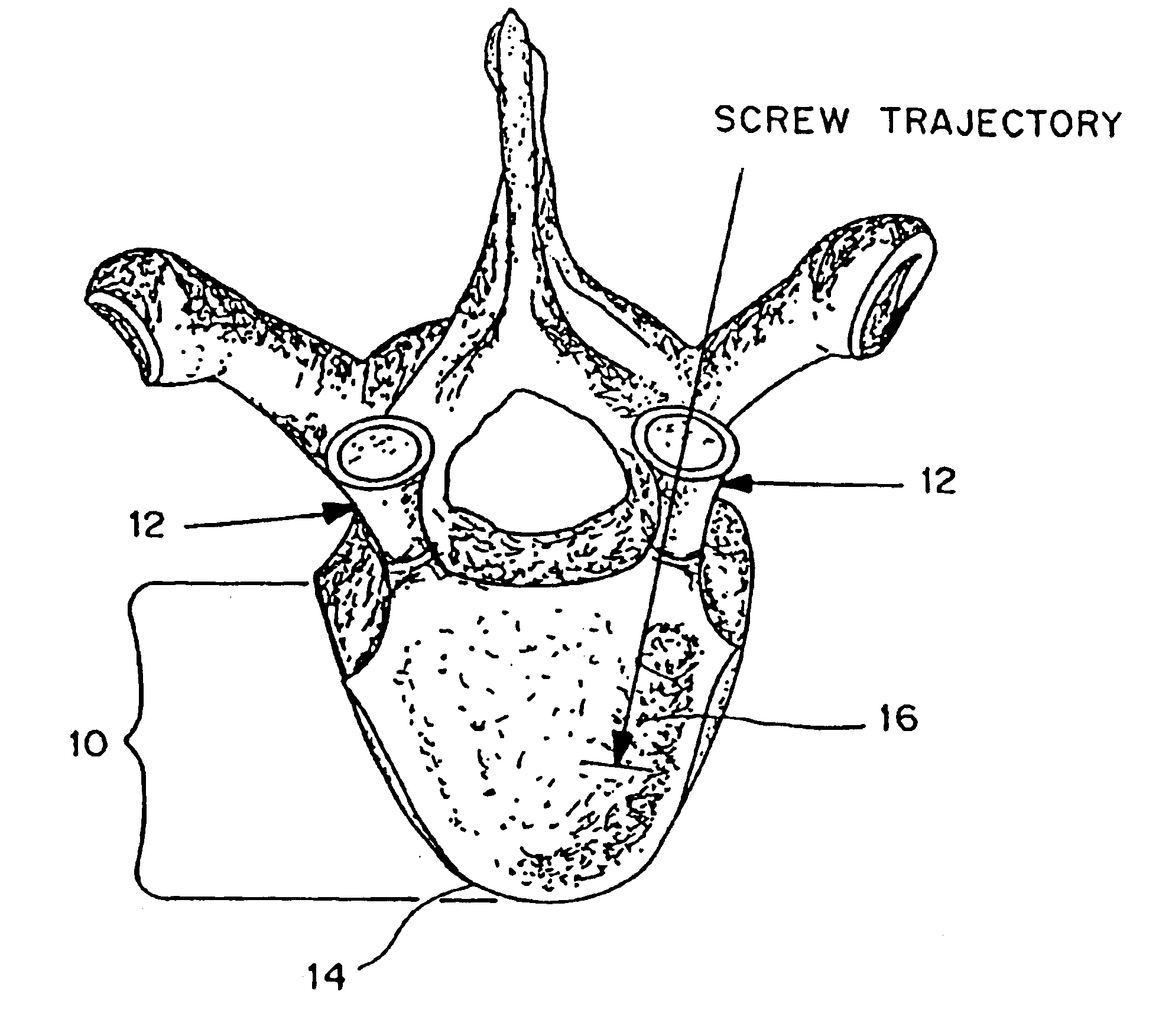
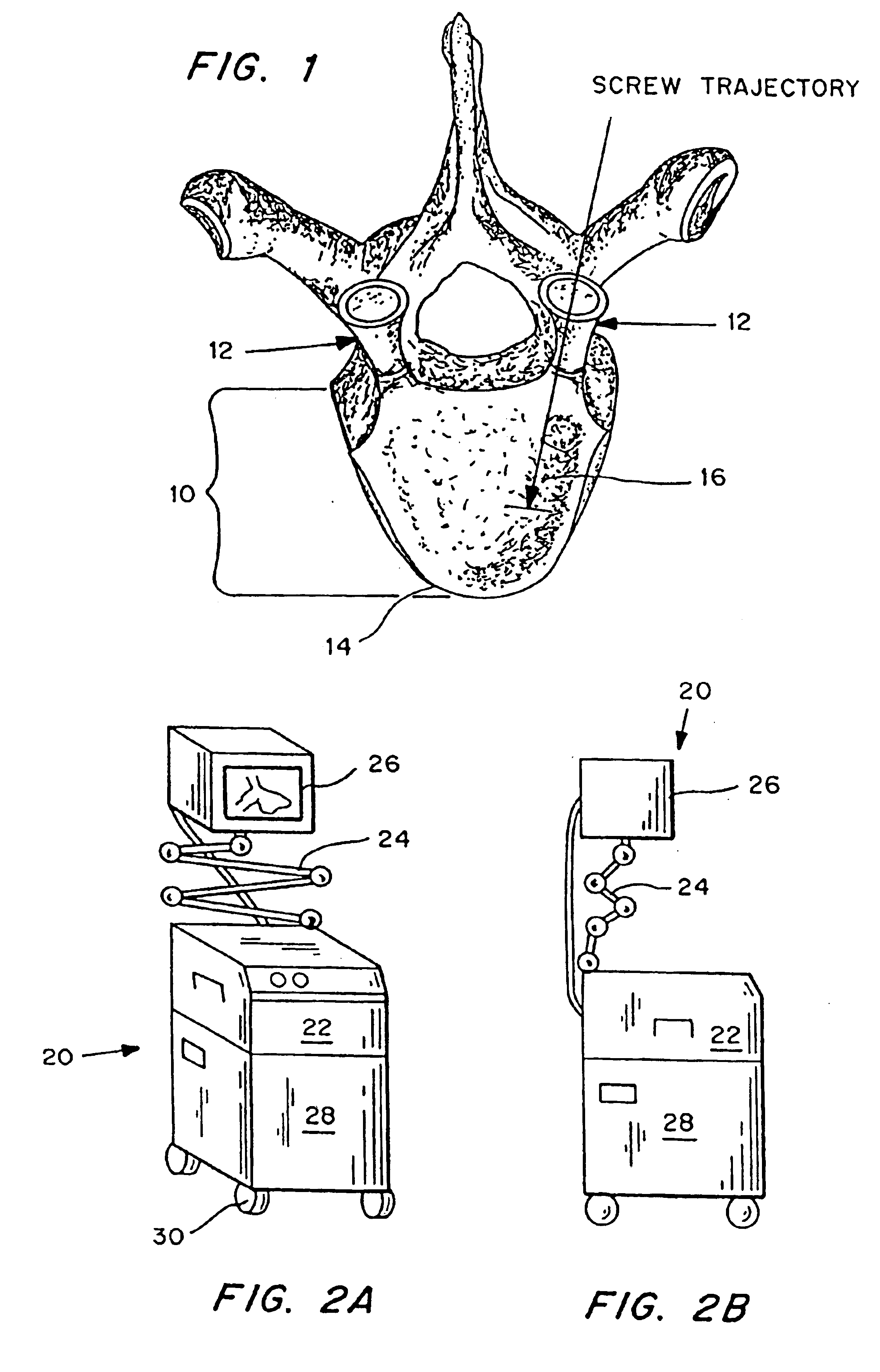
BACKGROUND OF THE INVENTIONThe present invention is generally in the field of methods and devices for surgical placement of implants, especially into bone.Surgical implantation of devices such as screws, pins, and other medical implants into bone is frequently the only means to safely immobilize the bone. Typically, this is done by passing a probe through the cortical bone, the dense, hard bone on the outside of bony structures, and into the cancellous bone, the soft, compliant spongy bone on the inside of the bone.As shown in FIG. 1, the relevant structures are the pedicles 12 and vertebral body 10. These structures are comprised of two types of bone: cortical 14 and cancellous 16. Cortical bone is the dense, hard bone covering the illustrated structures. Cancellous bone, commonly referred to as “spongy bone” is “soft” and compliant and provides the inner core for these structures.Surgeons exploit the difference in these two bone types during pedicular cannulation. When passing a b...

Claims


Application Information

Patent Timeline
Patent Type & Authority Patents(United States)
IPC IPC(8): A61B17/16A61B8/08A61B17/17A61B19/00A61B17/58A61B8/12
CPCA61B8/0841A61B8/0875A61B8/12A61B8/463A61B17/1626A61B17/1671A61B8/0833A61B8/0858A61B19/46A61B17/1757A61B8/4405A61B90/06
Inventor GOODWIN, MARK R.
Owner CUTTING EDGE SURGICAL
Why Eureka
Eureka Blog
Learn More