Photodynamic therapy in selective cell inactivation in blood and treating immune dysfunction diseases

a selective cell inactivation and photodynamic therapy technology, applied in the field of photodynamic therapy, can solve the problems of uncertainty in the nature of the disease progress and not in a uniform fashion

Inactive Publication Date: 2005-02-01
QLT INC
View PDF14 Cites 49 Cited by
  • Summary
  • Abstract
  • Description
  • Claims
  • Application Information

AI Technical Summary

Benefits of technology

irradiating the subject with light absorbed by said photoactive compound at an i

Problems solved by technology

The immunodisturbances caused by the human immunodeficiency virus have been studied, but the nature of the progress of the disease

Method used


Image

  • Photodynamic therapy in selective cell inactivation in blood and treating immune dysfunction diseases
  • Photodynamic therapy in selective cell inactivation in blood and treating immune dysfunction diseases
  • Photodynamic therapy in selective cell inactivation in blood and treating immune dysfunction diseases

Examples


example 1

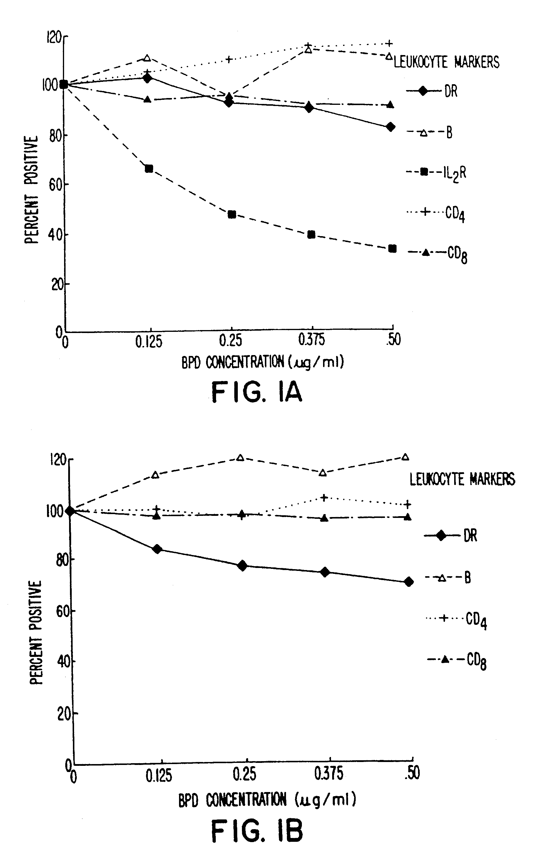
Response of Various Leukocytes to BPD and Irradiation

Blood was withdrawn from two patients who were shown to be infected with HIV. Whole blood from each patient was brought to varying concentration levels with BPD-MA and then irradiated with light at 10.8 J / cm2 using 690 nm light emitted from LEDs over a four-minute time period. The populations of various subsets of T-cells were evaluated by flow cytometry in comparison with untreated controls. The percentage of cells present was evaluated as a percentage of control and then plotted as a function of BPD-MA concentration.

The results obtained are shown in FIGS. 1A and 1B. As indicated, most cell populations remained relatively constant, including B-cells and CD4+ cells. Slight decreases were shown in CD8+ cells and DR+ cells. IL-2R+ cells showed a dramatic decrease for the patient results plotted in FIG. 1A, and this decrease was dependent on the dose of BPD-MA being used.

As shown in FIG. 1A, although IL-2R+ cells were dramatically de...

example 2

Whole human blood from an HIV patient was also subjected to treatment using various concentrations of BPD and various light intensities in a protocol similar to that set forth in Example 1. The effect on inactivation of cell-associated HIV was tested. The results obtained are shown in FIG. 4. As shown, intensities of 13 J / cm2 produced a dramatic inactivation of the virus at BPD concentrations of about 0.5 μg / ml or less. Lower intensities of radiation required higher concentrations of BPD to inactivate the virus completely.

example 3

The ability to inactivate free HIV (LAV-1 strain) in tissue culture of CEM cells was also tested. In the assay, LAV-1 stock was diluted into the tissue culture medium, and BPD was added at a concentration of either 0.25 μg / ml or 0.5 μg / ml. The media were incubated for one hour and exposed to three minutes of irradiation centered at 690 nm and at an intensity of 10.8 J / cm2. The media were then added to CEM cells, and the cells were assayed, using a standard p24 assay, after six days of culturing. The results were read in terms of pg / ml.

The results obtained are summarized below in Table 1. As shown, at dilutions of LAV that provided high levels of p24, treatment with BPD, a BPD,concentration of either 0.25 μg / ml or 0.5 μg / ml was capable of lowering substantially the level of p24 determined.

TABLE 1{overscore (x)} pg / ml (p 24)TREATMENTTREATMENTTREATMENTLAV-1No Drug.25 μg BPD.5 μg BPDDILUTIONno light3 min3 min10−2>61623.214.23 × 10−2>61618.214.55 × 10−2>61615.111.47 × 10−2>6168.97.69 × 1...


PUM

PropertyMeasurementUnit
Toxicityaaaaaaaaaa

Abstract

Multiple sclerosis and rheumatoid arthritis can be treated effectively using photodynamic therapy. In this protocol, a photoactive compound is administered, allowed to distribute in the effected subject, and the subject is then irradiated to activate the photoactive compound. Alternatively, the blood of a subject to be treated can be subjected to PDT extracorporeally. In the case of rheumatoid arthritis, localized treatment at the joints may also be employed.

Description

TECHNICAL FIELDThe invention relates to photodynamic methods for selectively depleting blood or bone marrow of cells important in the progress of certain diseases. More specifically, it concerns methods for selectively reducing the population of activated leukocytes in the blood or bone marrow of subjects with immune dysfunctional disorders, such as patients with autoimmune diseases or patients infected with HIV. Even more specifically, it concerns methods for treating specific autoimmune diseases such as multiple sclerosis and rheumatoid arthritis using photodynamic therapy.BACKGROUND ARTThe immunodisturbances caused by the human immunodeficiency virus have been studied, but the nature of the progress of the disease is still uncertain. However, it has been shown that HIV-infected humans have circulating cytotoxic T-lymphocytes that lyse uninfected CD4+ cells. Zarling et al., J. Immunol., 144:2992 (1990). It is also generally understood that infection with HIV, while depleting the l...

Claims


Application Information

Patent Timeline
IPC IPC(8): A01N25/00A61B10/00A61M37/00F21V9/16F21V9/00G01N21/01A61K31/40A61K31/409A61K31/46A61K31/555C07D487/16A61K41/00A61K47/00A61P29/00A61P31/18A61P37/00A61P37/06C07D487/22
CPCA61K31/40A61K31/409A61K41/0071Y10S514/885Y10S514/908Y10S514/825A61P29/00A61P31/18A61P37/00A61P37/06A61P43/00
Inventor LEVY, JULIA G.NORTH, JANICE
Owner QLT INC
Why Eureka
Try Eureka