
Method and system for magnetic resonance imaging

a magnetic resonance and imaging technology, applied in the field of magnetic resonance imaging, can solve the problems of time-consuming search for and tracking of interventional instruments, and achieve the effect of simplifying the tracking of objects

Inactive Publication Date: 2005-04-19
SIEMENS HEATHCARE GMBH
View PDF4 Cites 26 Cited by
  • Summary
  • Abstract
  • Description
  • Claims
  • Application Information

AI Technical Summary

Benefits of technology

[0009]An object of the present invention is to provide a method as well as a system for magnetic resonance imaging that enable the simplified tracking of an object located in a body channel, particularly a vessel, of a body region to be examined.
[0012]In the utilization of the present method, a prescribable region, particularly the center, of the respective MR tomogram is always situated in the segment of, for example, a vessel intersected by the trajectory. By the angling of the acquired slice calculated from the trajectory, an optimally large segment of the trajectory or of the appertaining vessel or body canal can be respectively recognized in the MR tomogram. This facilitates the locating and the tracking of the object. The angular attitude for each MR tomogram can be calculated, for example, via the tangent at the respective position along the trajectory or the position of the corresponding neighboring points on the trajectory. The position of the respective slice is therefore adapted to the course of the trajectory or of the vessel. The operator of the MR system thus need only specify the desired slice position either directly or via a corresponding position on an axis of the coordinate system of the MR system, and the MR tomogram thereby is optimally oriented for the tracking of the object situated in the body. Of course, a manual adaptation of the angular attitude or position of the tomogram remains possible.
[0017]In an embodiment of this MR system, a measurement device is additionally provided that is connected to the calculating unit or to the input unit, that measures an introduce length of an instrument introduced into the body region and provides this measurement to the calculating device and / or input device as position information. The measurement device can be fashioned as optical or mechanical device, for example as a slide resistor. In this way, the specification of the slice position at which the next MR tomogram should be acquired is not longer required but is automatically supplied by the measurement device.

Problems solved by technology

This procedure is also repeated after the locating of the intervertional instrument when the latter is moved forward in the framework of the minimally invasive intervention, The search for and the tracking of the interventional instrument are extremely time-consuming compared to a competing imaging technique wherein the tracking of the instrument ensues with conventional X-ray fluoroscopy.
A comparable problem arises in instances wherein an object, for example an endoscope or a probe, introduced into a body canal or tract is to be tracked with MR imaging.

Method used


Image

  • Method and system for magnetic resonance imaging
  • Method and system for magnetic resonance imaging
  • Method and system for magnetic resonance imaging

Examples


Embodiment Construction

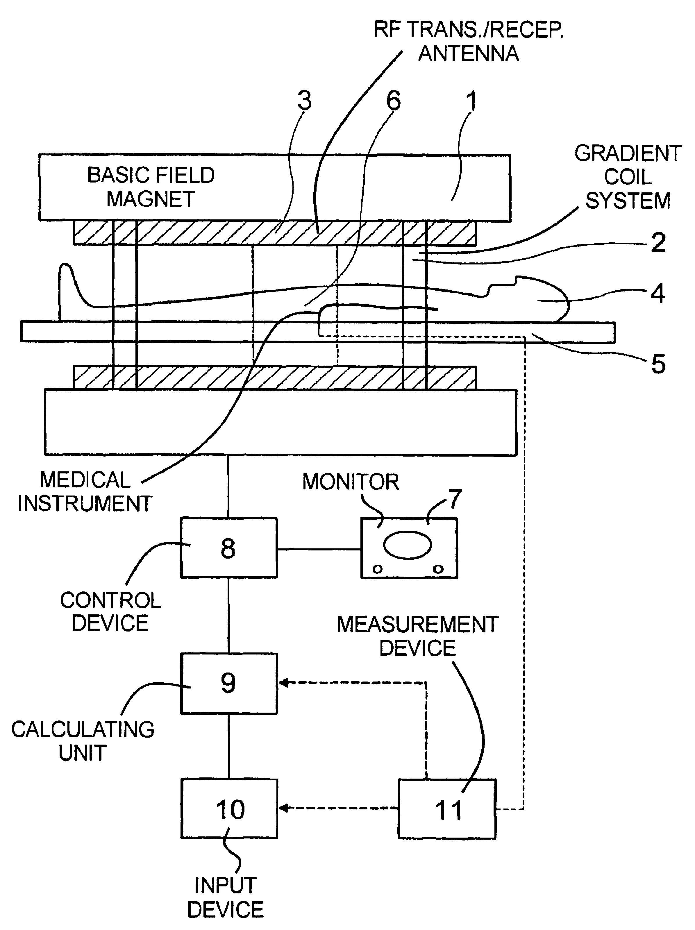
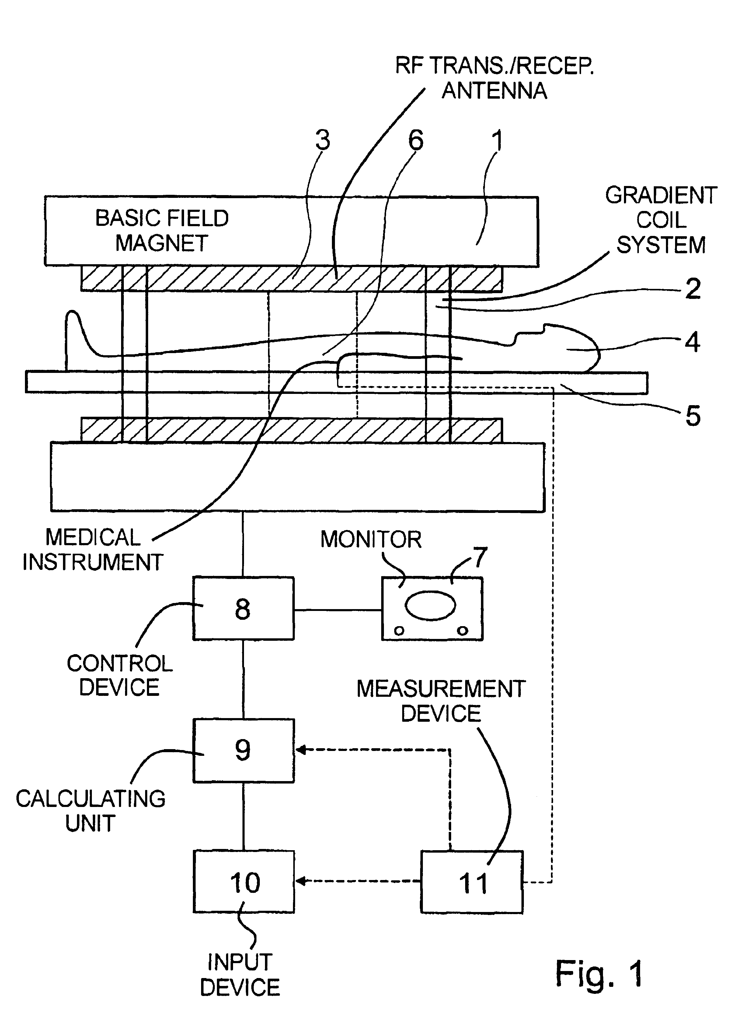
[0023]FIG. 1 schematically shows a section through a magnetic resonance tomography apparatus that can be utilized for the implementation of the present method. Only the basic component parts of the apparatus are shown in FIG. 1, namely a basic field magnet 1, a gradient coil system 2 and a radio-frequency transmission and reception antenna 3. A patient 4 also is shown on a patient bed 5, the patient 4 representing the examination subject. In the measurement, one or more radio-frequency pulses for generating magnetic resonance signals are radiated into the body of the person 4 via the radio-frequency transmission antenna 3, and the generated magnetic resonance signals are acquired and presented in the form of a two-dimensional MR tomogram or MIP image. With broken lines, FIG. 1 shows the body region 6 of the patient in which an introduced object, a stent in the present examples introduced by a schematically-indicated catheter 15, is to be tracked with MR imaging during the introducti...


PUM


Abstract

In a method and a system for magnetic resonance (MR) imaging, an MR exposure of a body region is implemented with an MR system, a trajectory within the body region is prescribed in a coordinate system of the MR system on the basis of the MR exposure, and MR tomograms within the body region at individual positions along the trajectory are automatically angled relative to the patient or magnet coordinate system dependent on a perspective pre-selected by an operator. This angling is calculated from the trajectory at the individual positions in the prescribed perspective such that a maximum segment of the trajectory is contained in each of the MR tomograms. The tracking of objects in body canals or vessels by means of MR imaging is considerably simplified with the method and the appertaining system.

Description

BACKGROUND OF THE INVENTION[0001]1. Field of the Invention[0002]The present invention is directed to a method for magnetic resonance (MR) imaging, wherein multiple MR tomograms at different positions within a body region to be examined are acquired in temporal succession and presented. The invention is also directed to an MR system for the implementation of such a method.[0003]2. Description of the Prior Art[0004]Magnetic resonance tomography is a known technology for acquiring images of the inside of the body of a living examination subject. For implementation of magnetic resonance tomography, a basic field magnet generative a static, relatively homogeneous basic magnetic field. During the acquisition of the MR tomograms, rapidly switched gradient fields that are generated by gradient coils are superimposed on this basic magnetic field. Radio-frequency pulses for triggering magnetic resonance signals are emitted into the examination subject with radio-frequency transmission antenna...

Claims


Application Information

Patent Timeline
IPC IPC(8): G01R33/54G01R33/28A61B5/055G01R33/32
CPCG01R33/4833G01R33/285A61B5/055
Inventor NITZ, WOLFGANG
Owner SIEMENS HEATHCARE GMBH
Learn More