CT detector reflector useful in detector scintillator array

a detector and reflector technology, applied in the field of computer tomograph (ct) imaging system, can solve the problems of reducing the reflectivity, reducing the light output of each photodiode detector cell, etc., and achieves the effects of less susceptible to x-ray damage, reducing cross talk, and reducing manufacturing costs

Inactive Publication Date: 2005-06-07
GE MEDICAL SYST GLOBAL TECH CO LLC
View PDF19 Cites 4 Cited by
  • Summary
  • Abstract
  • Description
  • Claims
  • Application Information

AI Technical Summary

Benefits of technology

[0007]There is therefore provided in one embodiment of this invention, a computed tomograph (CT) imaging system having a rotating gantry, a radiation source, a detector array on the rotating gantry and configured to detect radiation from the radiation source and the detector array. The detector array includes a photosensor array and an array of scintillators optically coupled to the photosensor array and a thermoplastic encased reflective film between and in front of the scintillators of the scintillator array. The reflector is thin and is less susceptible to x-ray damage.
[0008]These and other embodiments of the invention provide various combinations of additional advantages, including lower manufacturing cost due to use of a lamination reflector fabrication process, and lower cross talk.

Problems solved by technology

Such cast reflectors are sometimes damaged by X-ray exposure which causes color center formation, reduces reflectivity and causes lower light output from each photodiode detector cell.

Method used


Image

  • CT detector reflector useful in detector scintillator array
  • CT detector reflector useful in detector scintillator array
  • CT detector reflector useful in detector scintillator array

Examples


Embodiment Construction

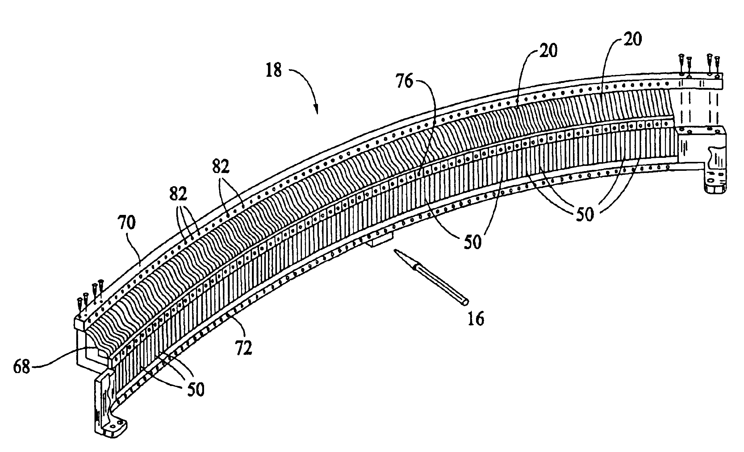
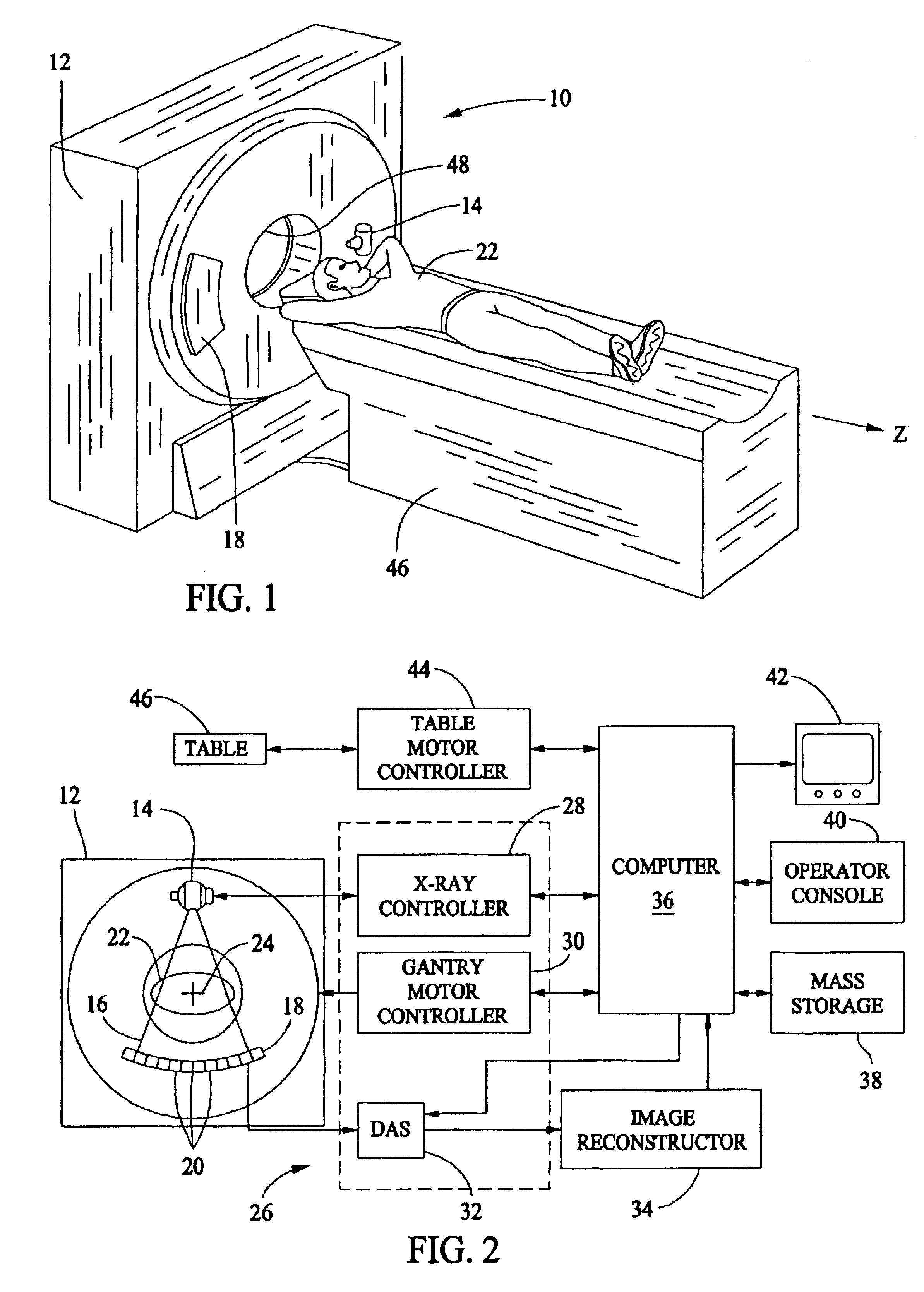
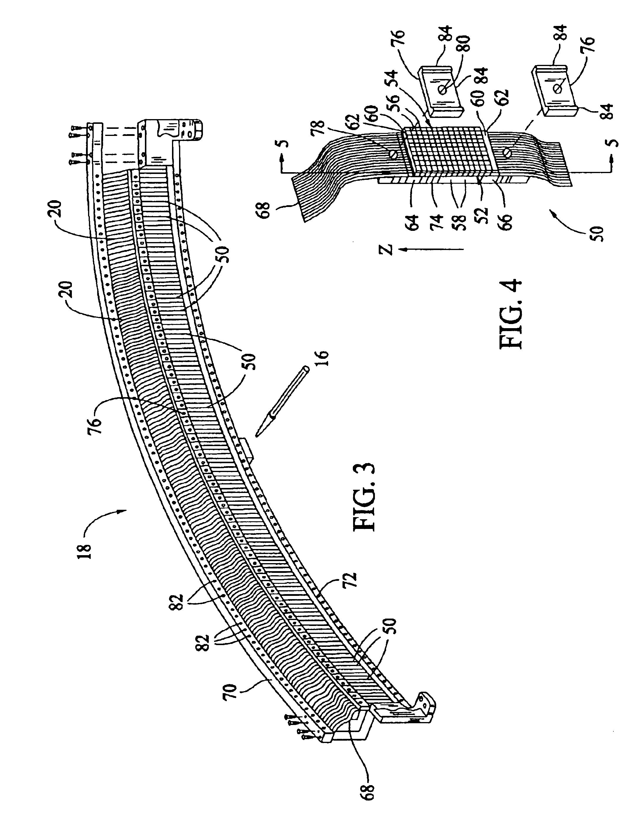
[0014]Referring to FIGS. 1 and 2, a computed tomography (CT) imaging system 10 is shown as including a gantry 12 representative of a “third generation” CT scanner. Gantry 12 has an x-ray source 14 that projects a beam of x-rays 16 toward a detector array 18 on the opposite side of gantry 12. Detector array 18 is formed by detector elements 20 which together sense the projected x-rays that pass through an object 22, for example a medical patient. Each detector element 20 produces an electrical signal that represents the intensity of an impinging x-ray beam and hence the attenuation of the beam as it passes through patient 22. During a scan to acquire x-ray projection data, gantry 12 and the components mounted thereon rotate about a center of rotation 24. Detector array 18 may be fabricated in a single slice or multi-slice configuration. In a multi-slice configuration, detector array 18 has a plurality of rows of detector elements 20, only one of which is shown in FIG. 2.

[0015]Rotatio...


PUM


Abstract

A method for making a detector array comprising a photosensor and scintillators, including placing a thermoplastically encased reflective film between scintillators of a scintillator array and optically coupling the scintillators with the photosensor.

Description

CROSS REFERENCE TO RELATED APPLICATIONS[0001]This application is a Continuation In Part of U.S. patent application Ser. No. 09 / 751,872 filed Dec. 29, 2000 now abandoned.BACKGROUND OF THE INVENTION[0002]This invention relates generally to a computer tomograph (CT) imaging system and more particularly to a CT detector module and reflector useful therewith and to methods for preparing and using the detector module and reflector.[0003]In at least one known computed tomography (CT) imaging system configuration, an x-ray source projects a fan-shaped beam which is collimated to lie within an X-Y plane of a Cartesian coordinate system and generally referred to as the “imaging plane”. The x-ray beam passes through the object being imaged, such as a patient. The beam, after being attenuated by the object, impinges upon an array of radiation detectors. The intensity of the attenuated beam radiation received at the detector array is dependent upon the attenuation of the x-ray beam by the object...

Claims


Application Information

Patent Timeline
Patent Type & Authority Patents(United States)
IPC IPC(8): G01T1/20G01T1/00A61B6/03
CPCG01T1/2002
Inventor HOFFMAN, DAVID M.
Owner GE MEDICAL SYST GLOBAL TECH CO LLC
Why Eureka
Try Eureka