Method and apparatus for evaluation of contrast agent uptake based on derived parametric images

a contrast agent and parametric image technology, applied in the field of medical imaging arts, can solve the problems of inconsistent diagnosis, cardiac disease is a leading health problem, and myocardial infarction, and achieve optimal efficiency, maximize the probability of acquiring high-quality functionals, and improve clinical work flow

Inactive Publication Date: 2005-06-07
KONINKLIJKE PHILIPS ELECTRONICS NV
View PDF7 Cites 74 Cited by
  • Summary
  • Abstract
  • Description
  • Claims
  • Application Information

AI Technical Summary

Benefits of technology

[0017]One advantage of the present invention is that it takes advantage of the complementary strengths of the BOLD and FPM imaging techniques to produce an improved clinical work flow.
[0018]Another advantage of the present invention resides in an optimization of slice positioning and orientation for transient contrast agent-enhanced imaging.
[0019]Another advantage of the present invention is that it provides for obtaining piloting information such as an optimized imaging region of interest, number of slices, slice location, slice orientation, slice thickness, imaging field of view, and the like, prior to performing time critical imaging of a transient contrast agent distribution in the optimized region of interest.
[0020]Another advantage of the present invention is that it maximizes the probability of acquiring high quality functional cardiac MRI images.
[0021]Yet another advantage of the present invention is that it makes optimally efficient use of limited clinical MRI facility time resources.
[0022]Numerous additional advantages and benefits of the present invention will become apparent to those of ordinary skill in the art upon reading the following detailed description of the preferred embodiment.

Problems solved by technology

It is well known that cardiac diseases are a leading health problem in the United States at the present time.
Myocardial infarctions, popularly known as heart attacks, are a leading cause of death.
However, in spite of its prevalence, diagnosis is inconsistent.
The incipient forms of cardiac disease can produce chest pains, excessive weariness, and other symptoms which usually only manifest during periods of exercise or other strenuous exertion.
Conversely, if left untreated the incipient forms often develop into more serious coronary diseases that lead to major heart attacks or other life threatening medical conditions.
The early coronary vascular blockages typically occur in small blood vessels which do not resolve well in conventional medical imaging techniques such as magnetic resonance imaging (MRI) or multi-slice computed tomography (CT).
The vessels involved are too small and too numerous to practically investigate using vascular catherization diagnostic techniques.
Catheterization procedures are also expensive and carry a higher degree of medical risk, further reducing their attractiveness for diagnosing ambiguous cases.
It is critical that the area imaged during this short time includes the cardiac region affected by the vascular blockage or blockages, and a failure to do so will typically result in a failed test and potentially incorrect medical diagnosis.
However, because of the limited available imaging time, tradeoffs are made between the volume coverage and the resolution with the result that full imaging coverage of the entire heart with satisfactory resolution is seldom achievable in FPM.
Once the Gd is substantially absorbed, however, it typically takes on the order of tens of minutes to hours for the Gd agent to be fully removed from the blood by the kidneys.
The slow removal of Gd from heart tissue makes it difficult to reliably repeat high-quality cardiac FPM imaging using more than one Gd bolus injection in a single imaging session due to residual contamination from previous Gd boluses.
In the past, the MRI operator attempting to acquire FPM images for evaluating myocardial function and tissue viability has had very limited prior knowledge about appropriate imaging conditions.
Due to time constraints, a more complete spatial mapping of the heart, requiring on the order of nine slices to fully cover the entire heart with adequate slice resolution, is not practical during FPM imaging.
Increased speed through the use of higher gradient-slew rates may not be permissible due to safety considerations.
The slice selection is made without significant foreknowledge of the defect to be imaged, and so a strong possibility exists that the selected several slices will not optimally intersect the unknown defect.
Furthermore, the delay between administering the Gd and subsequent removal by the kidneys is variable on the order of tens of minutes to hours.
Such long and uncertain delays can be problematic in a clinical environment where the MRI facility is operated on a tight schedule.
In some cases, concern for patient safety or other issues may preclude application of the stress agent.

Method used


Image

  • Method and apparatus for evaluation of contrast agent uptake based on derived parametric images
  • Method and apparatus for evaluation of contrast agent uptake based on derived parametric images
  • Method and apparatus for evaluation of contrast agent uptake based on derived parametric images

Examples


Embodiment Construction

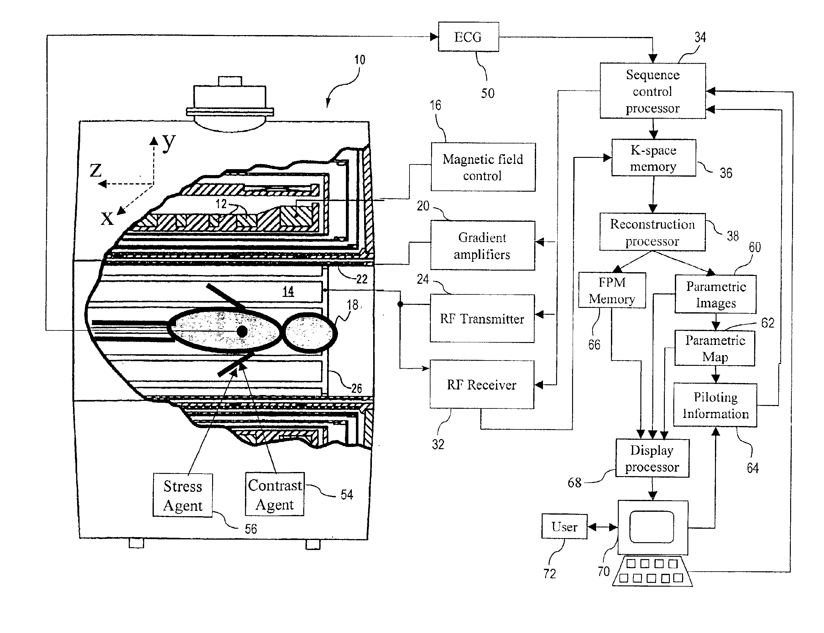
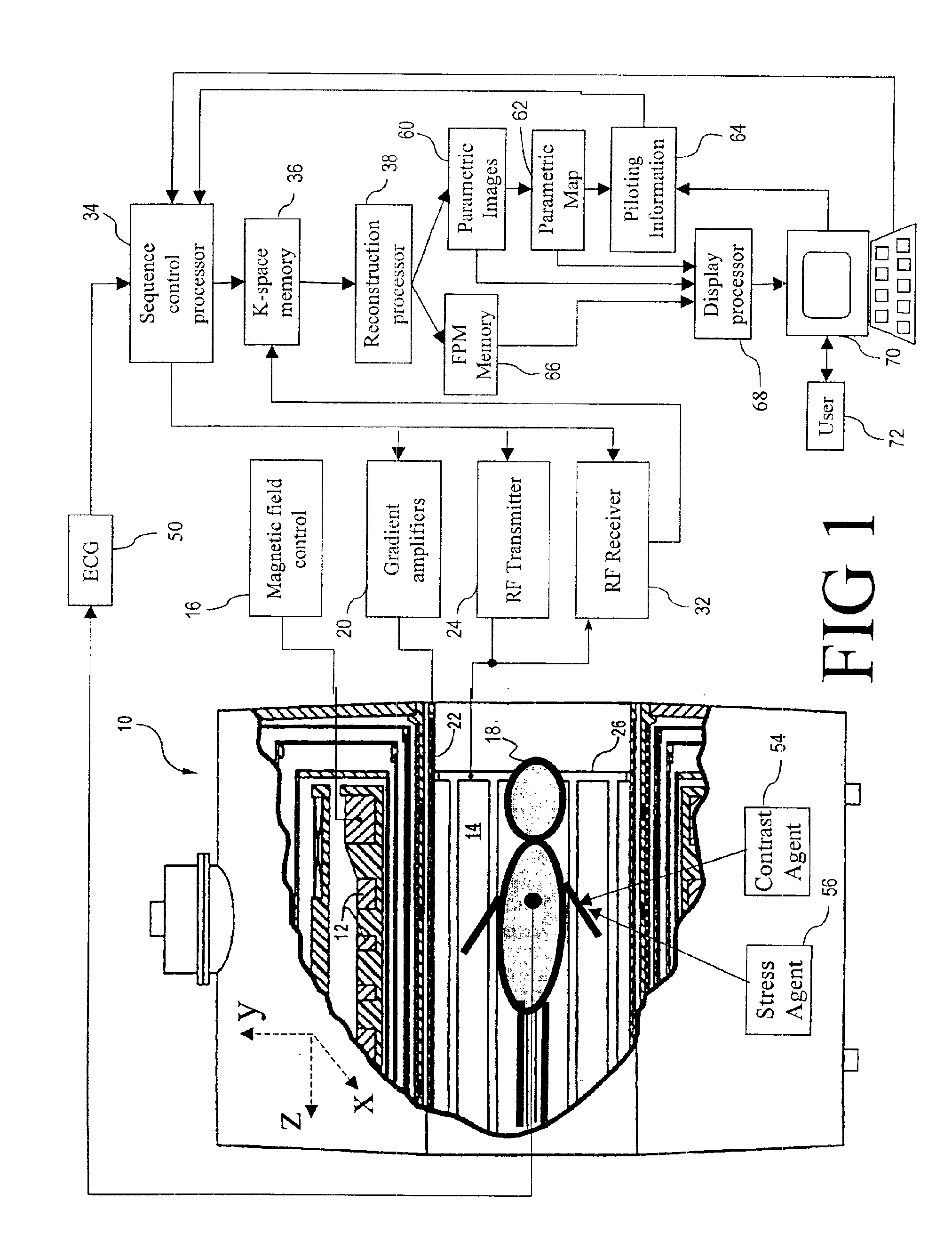
[0028]With reference to FIG. 1, a magnetic resonance imaging (MRI) scanner 10 includes superconducting or resistive magnets 12 that create a substantially uniform, temporally constant main magnetic field B0 along a z-axis through an examination region 14. Although a bore-type magnet is illustrated in FIG. 1, the present invention is equally applicable to open magnet systems and other known types of MRI scanners. The magnets 12 are controlled by a main magnetic field control 16. Imaging is conducted by executing a magnetic resonance (MR) sequence with the subject being imaged, e.g. a patient 18, placed with his or her heart or other region of interest within the examination region 14. Typically, the region of interest is placed at the isocenter.

[0029]The magnetic resonance sequence entails a series of RF and magnetic field gradient pulses that are applied to the subject to invert or excite magnetic spins, induce magnetic resonance, refocus magnetic resonance, manipulate magnetic reso...


PUM


Abstract

A magnetic resonance imaging (MRI) apparatus (10) acquires a plurality of parametric images (60) with at least one varying imaging parameter. A parametric map (62) is constructed from the plurality of parametric images (60). At least one pilot parameter (64) is identified from at least the parametric map (62). The at least one identified pilot parameter (64) includes at least a volume of interest for a diagnostic image. A contrast agent (54) is administered to the patient (18). The identified volume of interest is imaged during influx of the administered contrast agent (54) into the identified volume of interest. The imaging uses the at least one identified pilot parameter (64).

Description

BACKGROUND OF THE INVENTION[0001]The present invention relates to the medical imaging arts. It particularly relates to the assessment by magnetic resonance imaging (MRI) of coronary disease, heart tissue viability, and cardiac function, and will be described with particular reference thereto. However, the invention will also find application in other contrast-enhanced MRI imaging of various organs, such as the kidney, brain, and liver, as well as in other imaging modalities such as computed tomography (CT).[0002]It is well known that cardiac diseases are a leading health problem in the United States at the present time. Myocardial infarctions, popularly known as heart attacks, are a leading cause of death. However, in spite of its prevalence, diagnosis is inconsistent. More than half of the individuals who die of a heart attack do not exhibit previously recorded symptoms.[0003]Although often undiagnosed, many catastrophic myocardial infarctions are preceded by a prolonged period of ...

Claims


Application Information

Patent Timeline
Patent Type & Authority Patents(United States)
IPC IPC(8): A61B5/055G01R33/28G01R33/48G01R33/54
CPCA61B5/055A61B5/7292A61B5/7289
Inventor WU, DEE H.OBERRECHT, SARA M.HAGEY, SARAPRIATNA, AGUSFOXALL, DAVID L.
Owner KONINKLIJKE PHILIPS ELECTRONICS NV
Why Eureka
Try Eureka