

Real time measurement of cellular responses
a cellular response and real-time measurement technology, applied in the field of real-time observation of cellular proliferation, drug sensitivity, cell cycle position, etc., can solve the problems of cell death, dual labeled cells used,
- Summary
- Abstract
- Description
- Claims
- Application Information
AI Technical Summary
Benefits of technology
Problems solved by technology
Method used
Image

Examples
preparation b
Production of Histone H2B-GFP Vector
[0059]For histone H2B-GFP retrovirus production, the histone H2B gene was kindly provided by professor Geoff Wahl (Salk Institute). This gene has no stop codon, enabling the ligation of the H2B gene to the 5′ coding region of the Aequoria victoria EGFP gene (Clontech) 24). Then this histone H2B-GFP fusion gene was inserted at the Hind III / Cal I site of the pLHCX (Clontech) that has the hygromycin resistant gene. To establish a clone producing high amounts of a histone H2B-GFP retroviral vector, the pLHCX histone H2B-GFP plasmid was transfected to PT67 cells by same method for PT67-DsRed2. The transfected cells were cultured in the presence of 200-400 μg / ml of hygromycin (Life Technologies) for fifteen days and finally PT67-histone H2B-GFP cells were established. In some cases, after 15 days of drug selection, surviving colonies were checked under fluorescence microscopy and GFP or RFP-positive colonies were isolated. Several clones were selected a...
example 1
Human Prostate Cancer Cells-Expressing Histone H2B-GFP in the Nucleus and pLNC DsRed in the Cytoplasm
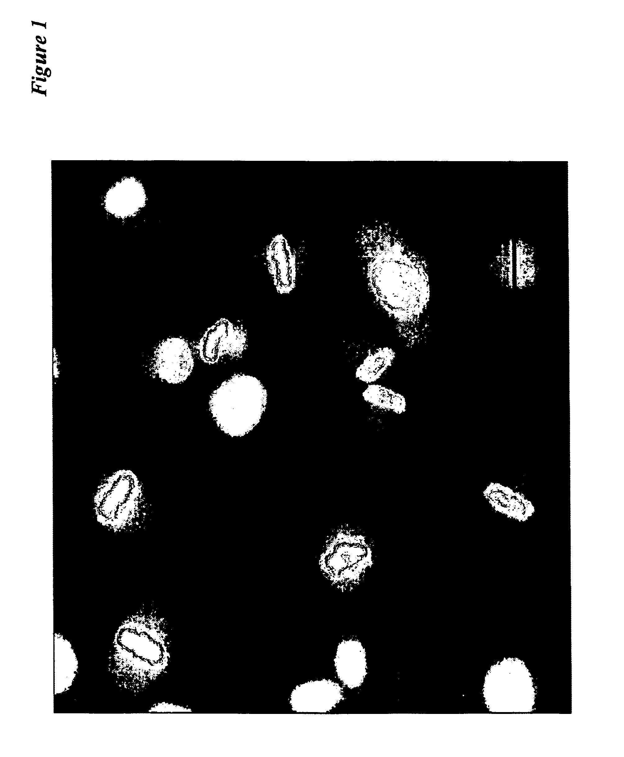
[0060]Dual color PC-3 cells were isolated that express GFP exclusively in the nucleus due to fusion of GFP with histone H2B (21) and express RFP exclusively in the cytoplasm. These cells demonstrate the feasibility of dual color imaging of live prostate cancer cells.
Step I: Preparation of DsRed-Expressing Cells.
[0061]PC-3 cells were transformed with pLNC DsRed-2 which is produced from PT67 packaging cells. The DsRed-s expression in the PC-3 cells was monitored under fluorescence microscopy. Selection was with increasing amounts of G418.
Step II: Preparation of Dual Labeled H2B GFP and DsRed-2 Cells.
[0062]The DsRed-2 PC-3 cells were transfected with pLHC H2B-GFP DNA using LipofectAMINE Plus™. After 24 hours incubation, the H2B GFP and DsRed-2-expressing cells were selected by increasing amounts of both hygromycin and G418.
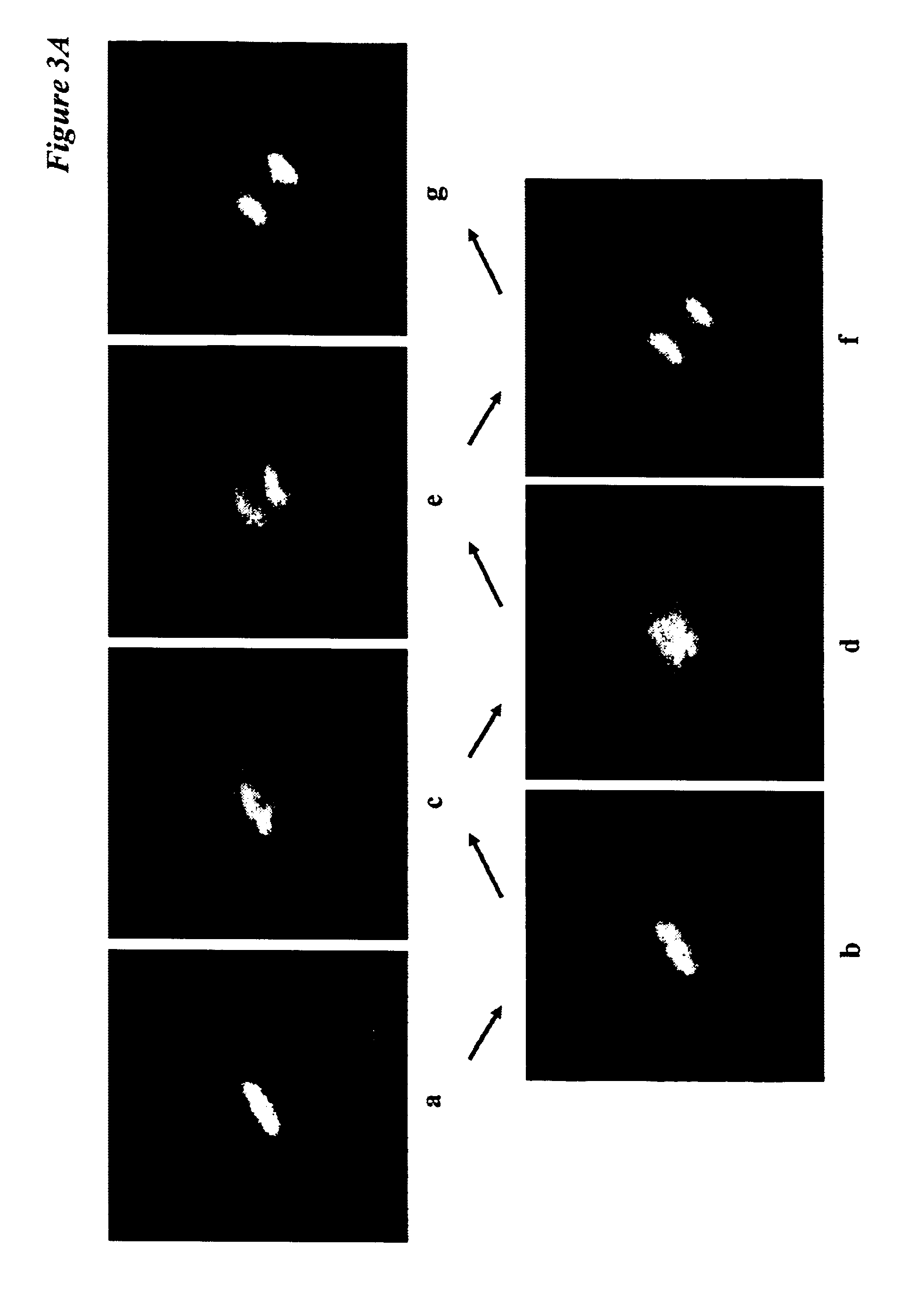
[0063]Cell-cycle position in living cells is analyzed by the are...
example 2
RFP and Histone H2B-GFP Gene Transduction of Fibrosarcoma Cells
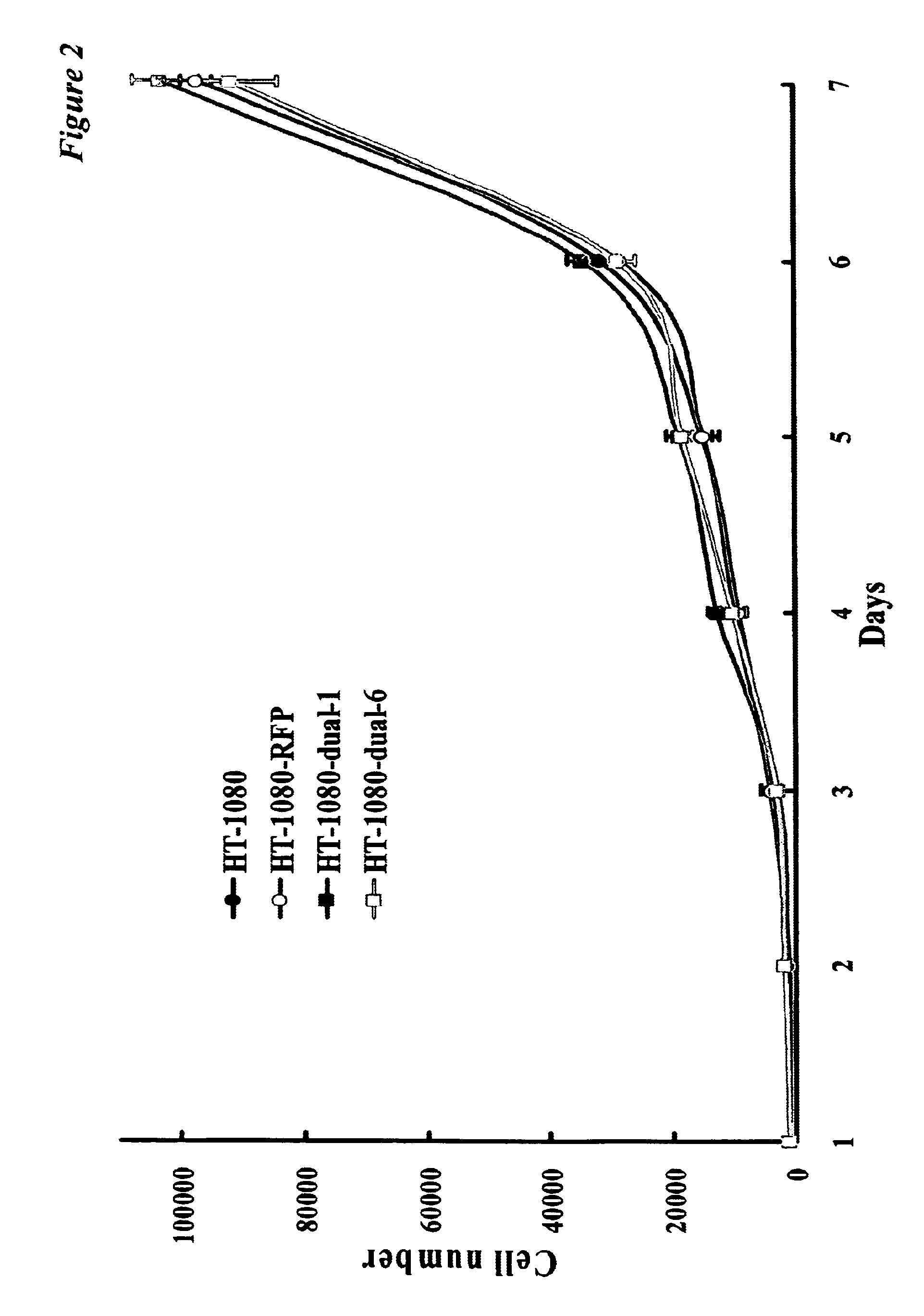
[0064]For RFP and histone H2B-GFP gene transduction, 70% confluent HT-1080 cells, derived from human fibrosarcoma and were purchased from American Type Culture Collection (Rockville, Md.). To establish dual-color cell, clones of HT-1080 expressing RFP in the cytoplasm (HT-1080-RFP), was established initially. Briefly, HT-1080 cells were incubated with a 1:1 precipitated mixture of retroviral supernatants of PT67-RFP cells and RPMI 1640 (Mediatech, Inc., Herndon, Va.) containing 10% fetal bovine serum for 72 hours. Fresh medium was replenished at this time. Cells were harvested by trypsin / EDTA 72 hours post-transduction and subcultured at a ratio of 1:15 into selective medium, which contained 200 μg / ml of G418. The level of G418 was increased stepwise up to 800 μg / ml. HT-1080-RFP were isolated with cloning cylinders (Bel-Art Products, Pequannock, N.J.) using trypsin / EDTA and amplified by conventional culture methods.
[0065...
PUM
| Property | Measurement | Unit |
|---|---|---|
| Time | aaaaa | aaaaa |
| Color | aaaaa | aaaaa |
| Ratio | aaaaa | aaaaa |
Abstract
Description
Claims
Application Information