Biocompatible form and method of fabrication

a biocompatible and fabrication technology, applied in the field of bone implants, can solve the problems of difficult restoration, unappealing aesthetically, loss of interproximal crestal alveolar bone, etc., and achieve the effect of accurately placing bone graft material and avoiding the problem of “black hole”

Inactive Publication Date: 2005-06-28
CAGENIX TECH
View PDF14 Cites 26 Cited by
  • Summary
  • Abstract
  • Description
  • Claims
  • Application Information

AI Technical Summary

Benefits of technology

[0013]In view of the foregoing needs, the present invention is directed to a biocompatible form, which may be permanently implanted in a patient's oral cavity for use in supporting bone graft material, and a method of fabricating the biocompatible form. The biocompatible form may be advantageously utilized in either intraosseous or subperiosteal applications and the configuration of the various embodiments of the biocompatible form permit the regeneration of the scalloped alveolar bone to normal skeletal contours, for either the maxilla or mandible, thereby permitting the restoration of missing teeth in their correct position. More particularly, the biocompatible form of the present invention permits the regeneration of normal interproximal bone and tissue for an aesthetic appearance, thereby avoiding the “black hole” problems associated with conventional bone grafting techniques. Additionally, the biocompatible form permits the regeneration of root prominence root bone contours and mylohyoid ridge bone contours which contribute to the proper placement of the replacement teeth. The various configurations of the biocompatible form of the present invention permit a dentist to accurately place the bone graft material and add structural support during healing to reproduce the alveolar bone contours required to place the replacement teeth and the associated implants in the correct position.

Problems solved by technology

When a person experiences a loss of teeth due to trauma or other circumstances, or has teeth with periodontal disease, there is often a loss of interproximal crestal alveolar bone.
This bone loss may also result in the loss of a person's interproximal or papillary oral tissue between the corresponding teeth and may cause a bone defect that is very unappealing aesthetically, and difficult to restore.
Without the proper regeneration of this bone defect, any replacement tooth is likely to be mal-positioned, out of proportion and shape and form and lack interproximal tissue for a natural appearance.
In the case of the mandible, the loss of teeth or periodontal disease may also result in a loss of mylohyoid ridge bone on either one or both sides of the mandible.
Loss of root prominence alveolar bone or mylohyoid ridge bone further complicates the ability of the dentist to properly regenerate the lost bone and makes it more likely that the artificial tooth will be improperly positioned since the corresponding dental implant or support structure is supported by the root prominence bone and, depending on the particular tooth, may also be supported by the mylohyoid ridge bone.
Known conventional graft techniques have failed to regenerate bone contours predictably and often result in placing grafted bone in locations where it is not needed, or regenerate bone that is over or under contoured.
This often causes the patient to have multiple tissue and bone surgeries to correct the contours of the first graft.
However, with the lack of bone in the atrophied maxilla or mandible, these criteria are impossible to accomplish and the results are poor with known techniques and devices.
No existing grafting technique attempts to restore the important interproximal scalloped bone contours that are critical to the proper placement of dental implants and aesthetics.
One of the main problems associated with conventional methods for replacing teeth with implants is the creation of a “black hole” defect between teeth.
This results from the lack of bone and tissue between the restored teeth and is very unnatural and is not aesthetically appealing to the patient.
Additional problems exist with known techniques associated with dental implants and bone grafts.

Method used


Image

  • Biocompatible form and method of fabrication
  • Biocompatible form and method of fabrication
  • Biocompatible form and method of fabrication

Examples


example 1

ous Implant

[0103]A patient with missing teeth requires restoration. A CAT scan of the area to be regenerated along with its surround arch form is performed. A computer-generated resin model of the existing bony contours and the remaining dental arch is fabricated. The model is mounted on articulator (Whipmix®, Lexington, Ky.) with the opposing arch. The missing teeth on the resin model are positioned in a normal anatomic position to determine the amount and contours of the bone to be regenerated. Wax or other medium is applied to the resin model to simulate the contours of the bone. The model is duplicated by conventional means. An intraosseous biocompatible form from the kit shown in FIG. 31 is selected that matches the patient. A biocompatible form 40 or 70 for a complete edentulous arch of the maxilla or mandible arch respectively, can be used or a biocompatible form such as 62, which is a portion of the full arch, can be used. The surgeon selects a biocompatible form that more c...

example 2

teal Implant

[0105]A patient with missing teeth requires restoration. A CAT scan of the area to be regenerated along with its surround arch form is performed. A computer-generated resin model of the existing bony contours and the remaining dental arch is fabricated. The model is mounted on articulator (Whipmix®, Lexington, Ky.) with the opposing arch. The missing teeth on the resin model are positioned in a normal anatomic position to determine the amount and contours of the bone to be regenerated. Wax or other medium is applied to the resin model to simulate the contours of the bone. The model is duplicated by conventional means. A subperiosteal biocompatible form from the kit shown in FIG. 31 is selected that matches the patient. The biocompatible form 40 is cut to precise form with titanium scissors and place on the duplicated model. The mesh can be recontoured with molding pliers to exactly fit the model and the desired regenerated contours. The biocompatible form 40 is sterilize...

example 3

efect Restoration

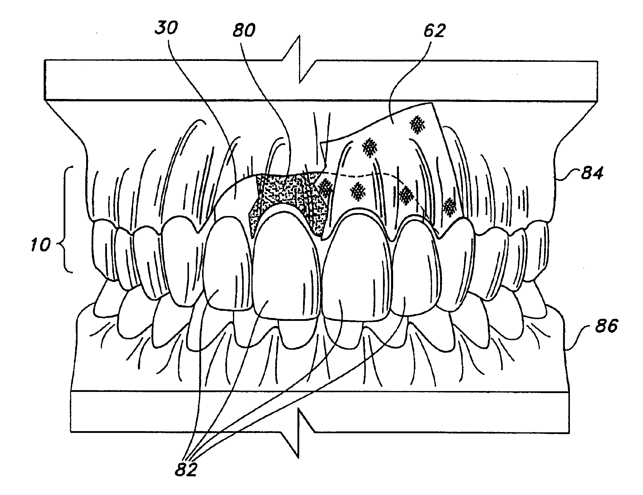
[0107]If a patient is missing palatal bone and several teeth due to surgery from a cancerous tumor, the procedures set out in Example 1 or 2 are repeated, but with the exception of a biocompatible form 60 having palatal area 61 is selected from the surgical kit 90.


PUM

PropertyMeasurementUnit
biocompatibleaaaaaaaaaa
areaaaaaaaaaaa
Biocompatibleaaaaaaaaaa

Abstract

A biocompatible form and a method for fabricating the implant are provided. The biocompatible form may be used to support bone graft material such as that used to reconstruct missing bone in a patient's oral cavity. The implant is fabricated from a biocompatible mesh, which may be made of titanium, a titanium alloy or fiber and is permanently implantable in the patient's oral cavity. The biocompatible form has an anatomical configuration which includes one or more portions conforming substantially to various alveolar bone contours which may include predetermined, human interproximal bone contours, root prominence bone contours and mylohyoid ridge bone contours. The biocompatible form may include a palatal section. The biocompatible form may also include one or more apertures for receiving a corresponding number of dental prostheses therethrough.

Description

CROSS-REFERENCE TO RELATED APPLICATIONS[0001]This is a division of U.S. patent application Ser. No. 10 / 012,652 filed Oct. 30, 2001 now U.S. Pat. No. 6,645,250 and entitled “Biocompatible Form and Method of Fabrication”. U.S. patent application Ser. No. 10 / 012,652 is hereby expressly incorporated by reference herein in its entirety.STATEMENT RE FEDERALLY SPONSORED RESEARCH OR DEVELOPMENT[0002]Not Applicable.REFERENCE TO A “MICROFICHE APPENDIX”[0003]Not Applicable.BACKGROUND OF THE INVENTION[0004]1. Field of the Invention[0005]The present invention relates generally to bone implants and, more particularly, to biocompatible forms for use in supporting bone graft material.[0006]2. Description of the Art[0007]When a person experiences a loss of teeth due to trauma or other circumstances, or has teeth with periodontal disease, there is often a loss of interproximal crestal alveolar bone. This bone loss may also result in the loss of a person's interproximal or papillary oral tissue betwee...

Claims


Application Information

Patent Timeline
Patent Type & Authority Patents(United States)
IPC IPC(8): A61C8/00
CPCA61C8/0006A61F2/2803A61F2/2846
Inventor SCHULTER, CARL W.
Owner CAGENIX TECH
Why Eureka
Try Eureka