Image pasting using geometry measurement and a flat-panel detector

a detector and geometry technology, applied in the field of xray images, can solve the problems of difficult handling, low image quality (iq) of many fs systems, and less accurate pasting imag

Inactive Publication Date: 2005-09-13
GE MEDICAL SYST GLOBAL TECH CO LLC
View PDF28 Cites 59 Cited by
  • Summary
  • Abstract
  • Description
  • Claims
  • Application Information

AI Technical Summary

Benefits of technology

[0015]Further, a system for creating a composite image from at least two images generated in response to x-ray radiation passing through first and second portions of human body is disclosed. The system comprises an x-ray source; a solid-state, flat-panel, x-ray detector including a first portion for converting x-rays to light, a second portion for converting light to information represented by electrons and a third portion configured to generate digital data representative of x-ray radiation based upon the information; a movement device coupled to the detector to facilitate relative movement between the detector and the human body along a straight path, such that the detector is positioned to generate first digital data representative of the first portion and positioned to generate second digital data representative of the second portion; a processor for processing the digital data to generate composite image date representative of the first and second portions of the body; a display coupled to the processor which presents a human viewable image generated based upon the image data; and a network for coupling the display to the processor. The first portion of the x-ray detector is preferably fabricated from cesium iodide, and the second portion preferably includes amorphous silicon and is preferably a photodiode / transistor array.
[0018]Also, a method for use with an x-ray device having an x-ray source and an x-ray detector, of determining a relative position between a first x-ray image and a second x-ray image which have been combined to form a pasted image is disclosed. The method comprises collecting a first image at a first relative position, collecting a second image at a second relative position, measuring a geometry of the first image, and using the geometry of the first image and a geometry of the second image to aid in determining the position of the first image relative to the position of the second image in the pasted image. In one embodiment, the geometry is measured by measuring the position of the detector, and in another embodiment, it is measured by measuring the position of the x-ray source. In still another embodiment, the position is measured by measuring the position of the detector relative to at least one point of reference.

Problems solved by technology

A problem with the current systems is that the digital images formed either need to be handled manually or do not give high quality images—making the pasted image less accurate.
Many FS systems have much lower image quality (IQ) than some detectors; are difficult to handle because several films must be manually placed into a cassette, and stored; have a lower dynamic range than some detectors; and cannot be post-processed, nor can they be quantitatively analyzed.
Typical CR systems tend to be difficult to handle because several PSP plates must be placed into a large, heavy, cumbersome, custom cassette; require extra time to insert and extract plates from the cassette; have a white band artifact at the region of overlap between the two plates due to attenuation; and have lower IQ than some detectors.
II systems tend to have poor IQ and introduce distortions such as pin-cushion effects.
The detector can also cause representation errors during image pasting (this has been referred to as the Pisa effect, or s-distortion), have anisotropic resolution across the field (the resolution is highest in the center of the detector, but decreases toward the periphery), may be sensitive to vibrations during movement causing additional image noise called “microphonic” noise.
Longer processing and pauses could mean a longer exam time, which in turn may cause an increase in patient movement artifacts.
Systems with a lower detective quantum efficiency (DQE) cause images to exhibit high noise levels.
High dose is especially undesirable in pediatric cases, and especially, as in image pasting, when the subject of interest must be exposed multiple times in order to get a full image.
Such systems do not work as efficiently as possible because there is no frame of reference which suggests a possible starting point for the overlap.

Method used


Image

  • Image pasting using geometry measurement and a flat-panel detector
  • Image pasting using geometry measurement and a flat-panel detector
  • Image pasting using geometry measurement and a flat-panel detector

Examples


Embodiment Construction

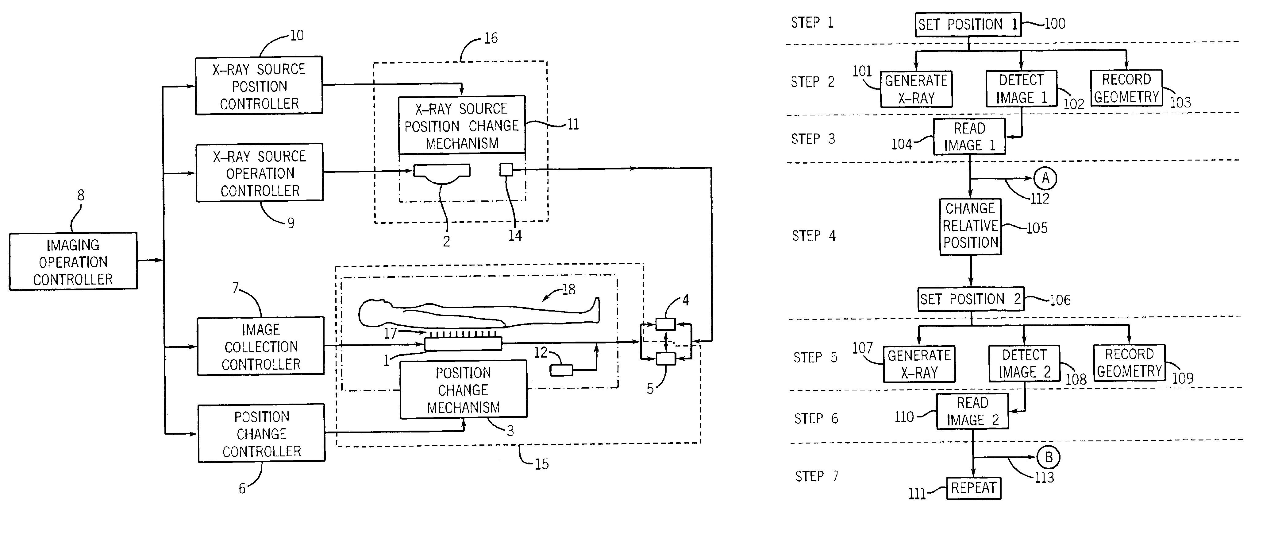
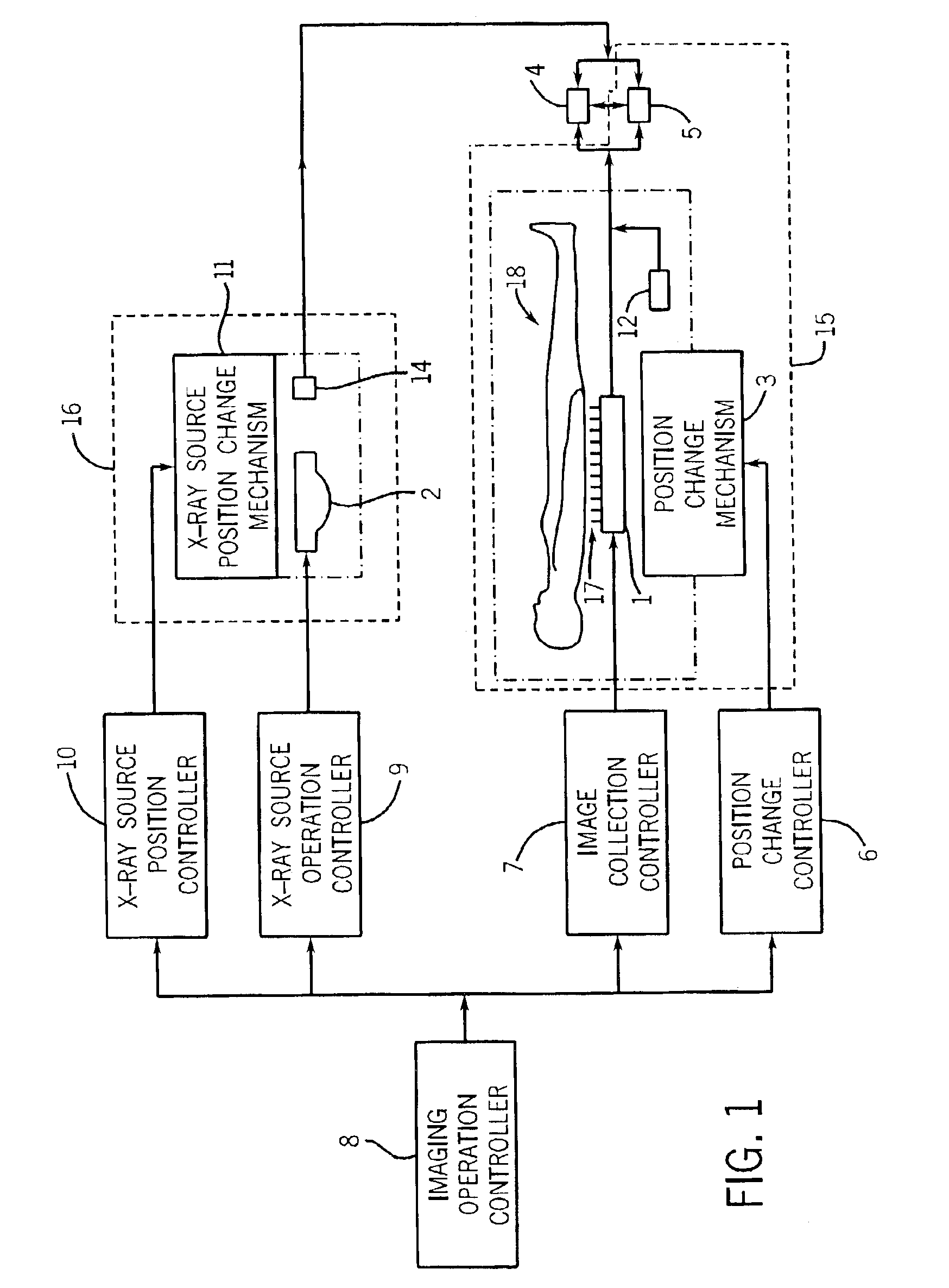
[0030]FIG. 1 is a schematic diagram showing the structure of an x-ray device according to one embodiment of the present invention. The x-ray device includes components such as an x-ray detector 1, an x-ray grid 17, an x-ray source 2, a position change mechanism 3, a processor 4, an image storage enabling unit 5, a position change controller 6, an image collection controller 7, an x-ray source controller 9, an x-ray source position controller 10, an x-ray source position change mechanism 11, an imaging operation controller 8, a positioner 12, and an inclinometer 14.

[0031]The x-ray device images a subject of interest 18. The subject of interest 18 can be any number of items where taking an x-ray image of the item is desired. Some typical subjects of interest are human patients to make diagnosis, sealed packages to determine contents, and welding joints to ensure a complete weld.

[0032]In FIG. 1, the subject of interest 18 is a human patient. The patient can stand in any position, e.g.,...


PUM


Abstract

A device for use in image pasting is described. The device includes a digital x-ray detector capable of automatic digital imaging without the use of an image intensifier; the detector preferably being a flat-panel detector. Additionally, an image pasting system using a solid-state detector is described. The system can connect the detected images to a display via a network (such as a WAN, a LAN, or the internet). Further, an image geometry measurement device for use in pasting x-ray images is disclosed. The geometry measurement device helps determine the relative position of two images to be used in image pasting. This information can be used alone, or in connection with an image pasting algorithm. Still further, methods of forming composite images are disclosed using a flat-panel detector and using the geometry of the images. The disclosed devices and systems can be integrated with other digital image pasting technology.

Description

FIELD OF THE INVENTION[0001]The present invention relates to collecting x-ray images generated using a digital detector for use in generating a composite image from a series of individual images.BACKGROUND OF THE INVENTION[0002]Image pasting, or the creation of a composite image, is usually accomplished by having a system for acquiring images with a total field-of-view larger than the detector field-of-view (FOV). For applications such as full-spine imaging or long-legs imaging, the total coverage of anatomy (e.g., 60-120 cm) exceeds that of most current detectors and film-screen systems. Modifications of the current systems need to be designed to allow imaging a total coverage larger than the detector FOV. A problem with the current systems is that the digital images formed either need to be handled manually or do not give high quality images—making the pasted image less accurate.[0003]Typical systems for image pasting use either: film-screen (FS) cassettes—either one long-film or ...

Claims


Application Information

Patent Timeline
Patent Type & Authority Patents(United States)
IPC IPC(8): A61B6/00
CPCA61B6/4233A61B6/4429A61B6/5241A61B6/547A61B6/563A61B6/4291
Inventor WARP, RICHARD J.BATTLE, VIANNEY P.KUMP, KENNETH S.METZ, STEPHEN W.HALSMER, MATTHEW A.UPPALURI, RENUKA
Owner GE MEDICAL SYST GLOBAL TECH CO LLC
Why Eureka
Try Eureka