Method for examining a specimen, and scanning microscope system

a scanning microscope and specimen technology, applied in the field of specimen examination, can solve the problems of living specimens, cement and lens elements are damaged irreversibly, and the use of ultraviolet illuminating light has the disadvantage, so as to improve the bleaching characteristics, the probability of multi-photon excitation is higher, and the power density is higher.

Inactive Publication Date: 2005-11-01
LEICA MICROSYSTEMS CMS GMBH
View PDF19 Cites 11 Cited by
  • Summary
  • Abstract
  • Description
  • Claims
  • Application Information

AI Technical Summary

Benefits of technology

[0022]The invention has the advantage that specimens marked with different fluorescent dyes can be examined in the context of simultaneous excitation and substantially improved bleaching characteristics.
[0023]In a further method step, the illuminating light is focused onto a subregion of the specimen, thereby resulting in a high

Problems solved by technology

The use of ultraviolet illuminating light has the disadvantage, especially for living specimens, of much more severe specimen damage.
With cemented optical components in particular, such as lens element groups in a microscope objective, illumination with ultraviolet light results in irreversible damage to the

Method used


Image

  • Method for examining a specimen, and scanning microscope system
  • Method for examining a specimen, and scanning microscope system
  • Method for examining a specimen, and scanning microscope system

Examples


Embodiment Construction

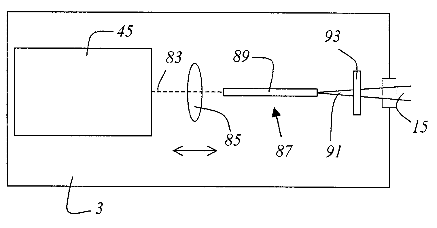
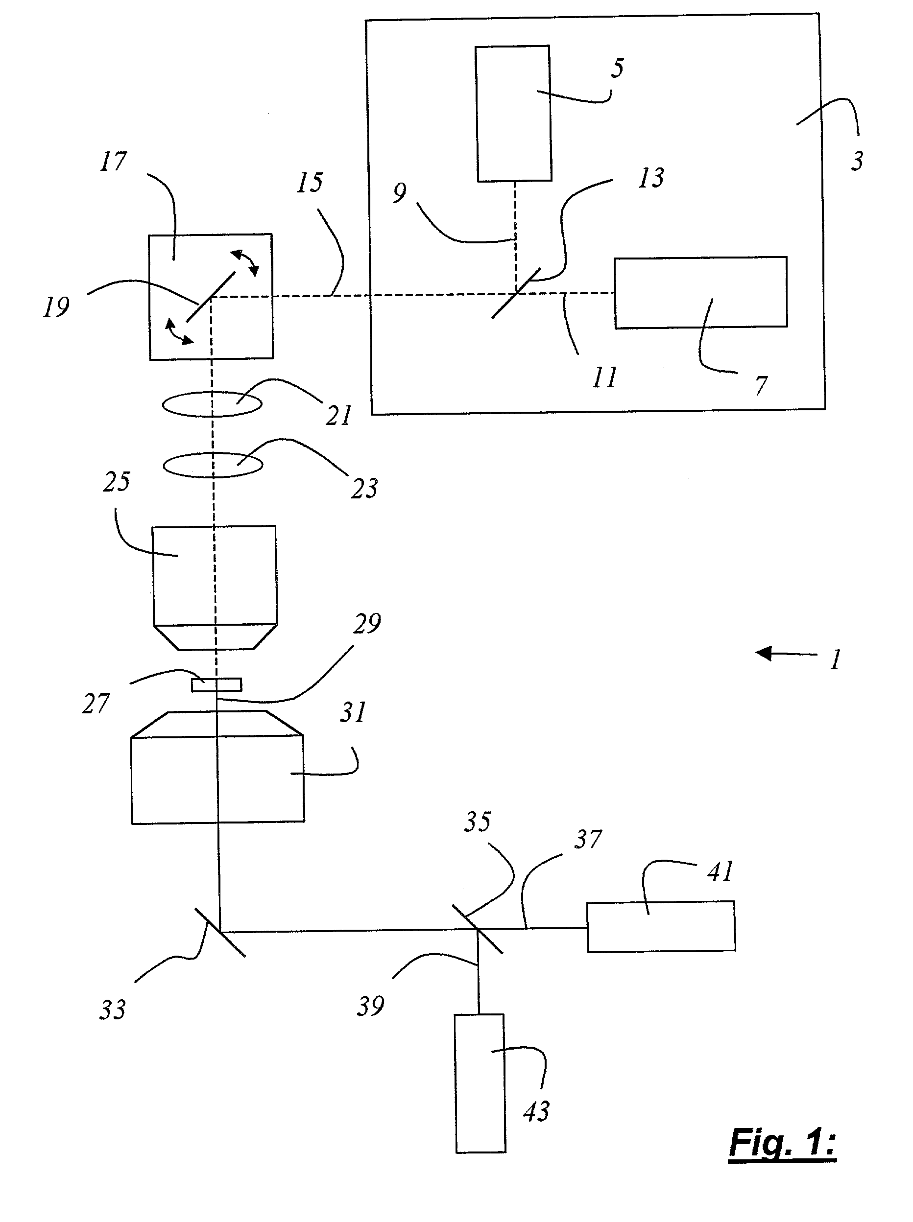
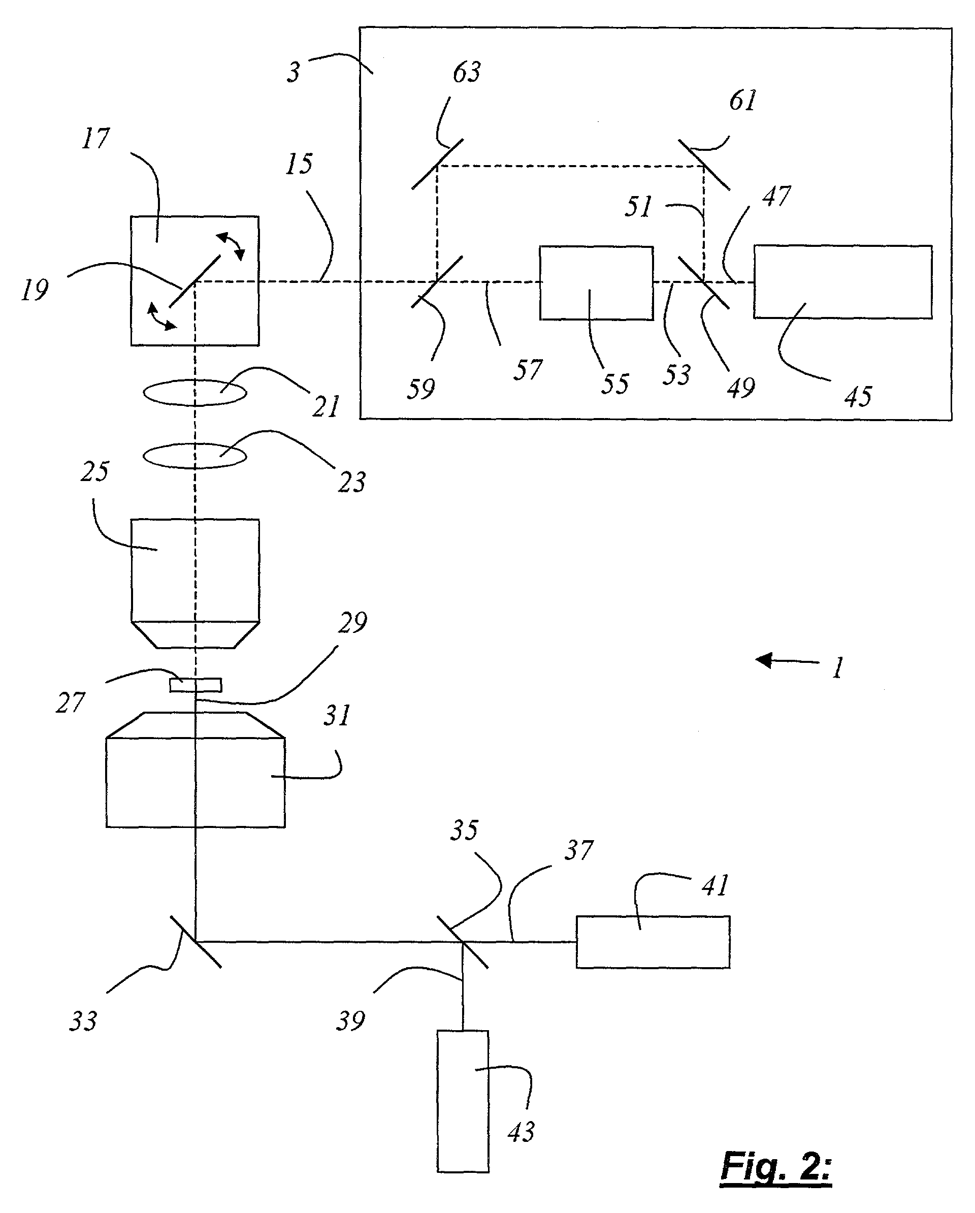
[0041]FIG. 1 schematically shows a scanning microscope system 1 according to the present invention having a light source 3 that contains a first and second laser 5, 7 embodied as mode-locked titanium / sapphire lasers. Light 9 emitted by first laser 5, which has a wavelength of 780 nm, and light 11 emitted by second laser 7, which has a wavelength of 960 nm, are combined with a dichroic beam splitter 13 into illuminating light 15. Illuminating light 15, shaped into a beam, arrives at a beam deflection device 17 that contains a gimbal-mounted mirror 19. By means of scanning optical system 21, tube optical system 23, and objective 25, illuminating light 15 that has been shaped into a beam is guided over or through specimen 27, which contains two fluorescent dyes that are optically excitable at 390 nm and 480 nm respectively. Detected light 29 proceeding from specimen 27 is collimated by a condenser 31 and reflected by a mirror 33 to a dichroic beam splitter 35 that divides the detected ...


PUM


Abstract

A method for examining a specimen (27) that exhibits at least two optical transition lines and is optically excitable at least with light of a first and light of a second wavelength is characterized by the step of illuminating the specimen (27) with illuminating light (15) that generates at least a multiple of the first wavelength and a multiple of the second wavelength; and by the step of detecting the detected light (29) proceeding from the specimen (27).
Also disclosed is a scanning microscope system (1) having at least one light source (3) that emits illuminating light (15) for illumination of a specimen (27), the specimen (27) exhibiting at least two optical transition lines and being optically excitable at least with light of a first and light of a second wavelength, having at least one detector (41, 43, 65, 77, 79) for detection of the detected light (29) proceeding from the specimen (27) and an objective (25) for focusing the illuminating light (15) onto a subregion of the specimen (27). The scanning microscope system is characterized in that the illuminating light (15) generates at least a multiple of the first wavelength and a multiple of the second wavelength.

Description

CROSS REFERENCE TO RELATED APPLICATIONS[0001]This application claims priority of the German patent application 101 20 425.6 which is incorporated by reference herein.FIELD OF THE INVENTION[0002]The invention concerns a method for examining a specimen that exhibits at least two optical transition lines and is optically excitable at least with light of a first and light of a second wavelength.[0003]The invention further concerns a scanning microscope system.BACKGROUND OF THE INVENTION[0004]For the examination of biological specimens, it has been usual for some time to prepare the specimen with optical markers, in particular with fluorescent dyes. Often, for example in the field of genetic investigations, several different fluorescent dyes which become attached to specific specimen constituents are introduced into the specimen. From the fluorescent properties of the prepared specimen it is possible, for example, to draw conclusions as to the nature and composition of the specimen, or a...

Claims


Application Information

Patent Timeline
IPC IPC(8): G02B21/00G01N21/64G01J3/30G01J3/36G02B21/06
CPCG01J3/36G01N21/6458G02B21/0048G02B21/0064G02B21/0076G01N2021/6441G02B2207/114
Inventor ENGELHARDT, JOHANNHOFFMANN, JUERGEN
Owner LEICA MICROSYSTEMS CMS GMBH
Why Eureka
Try Eureka