
Rapid response system for the detection and treatment of cardiac events

a rapid response and cardiac event technology, applied in the field of rapid response system for the detection and treatment of cardiac events, can solve the problems of life-threatening complications of heart disease, myocardial infarction, and no system that provides for early and automatic detection of acute myocardial infarction, and achieve the effect of increasing the heart ra

Inactive Publication Date: 2006-01-10
ANGEL MEDICAL SYST
View PDF17 Cites 207 Cited by
  • Summary
  • Abstract
  • Description
  • Claims
  • Application Information

AI Technical Summary

Benefits of technology

"The present invention is an implanted cardiosaver that can detect the occurrence of an acute myocardial infarction (AMI) and quickly alert the patient. The cardiosaver system includes an implanted cardiac monitor that can detect the AMI in less than five minutes after it occurs. The system also includes an external alarm system that can wake the patient and provide the patient with a display of their medical record and electrogram data for consideration by a medical practitioner. The network operation support system can also receive the patient's alarm and provide information to the medical practitioner about the patient's condition. The cardiosaver system is designed to minimize damage to the heart by detecting the AMI as early as possible and alerting the patient to seek medical care."

Problems solved by technology

A common and life-threatening complication of heart disease is myocardial infarction resulting from a thrombus that obstructs blood flow in one or more coronary arteries.
At this time, no system exists that provides for early and automatic detection of an acute myocardial infarction and for rapidly providing the patient with a prescribed dose of a thrombolytic medication.
However, no existing implantable pacemaker or defibrillator is also able to detect a partial or complete blockage of a coronary artery and warn the patient that this potentially fatal event is occurring.
Furthermore, no pacemaker or ICD is presently used with an external communication and action system that rapidly responds to acute myocardial infarction or reversible myocardial ischemia that is detected by specially designed circuitry in that implanted pacemaker or ICD.
Although anti-tachycardia pacemakers and ICDs can detect heart arrhythmias and respond with electrical stimulation, none are currently designed to be part of a rapid response system designed to inform the patient as to his condition and to facilitate the injection of an anti-arrhythmic drug with a minimum time delay.
However, the long term storage of any thrombolytic or anti-thrombogenic medication within a human patient can cause a deterioration of such drugs.
However, Zehender never discusses the treatment of acute myocardial infarction.
Furthermore, Zehender does not consider the problem of drug deterioration when such a drug is stored within an implanted device for later release when ischemia is detected.
Under such circumstances it would be highly undesirable to release a medication designed to treat the ischemia caused by a heart attack.
Furthermore, Zehender does not describe an external system to be used in conjunction with an implanted cardiosaver device.
Thus Zehender does not teach the use of an external alarm means or means to have a diagnostic center always on call for immediate diagnosis of a potentially fatal heart attack or arrhythmia.
Furthermore, Zehender does not teach an implanted drug port that can be used for rapidly providing a bolus of medication from an external source to be delivered into a patient having a heart attack by either the patient himself, a caretaker of the patient or by a paramedic from an ambulance.

Method used


Image

  • Rapid response system for the detection and treatment of cardiac events
  • Rapid response system for the detection and treatment of cardiac events
  • Rapid response system for the detection and treatment of cardiac events

Examples


Embodiment Construction

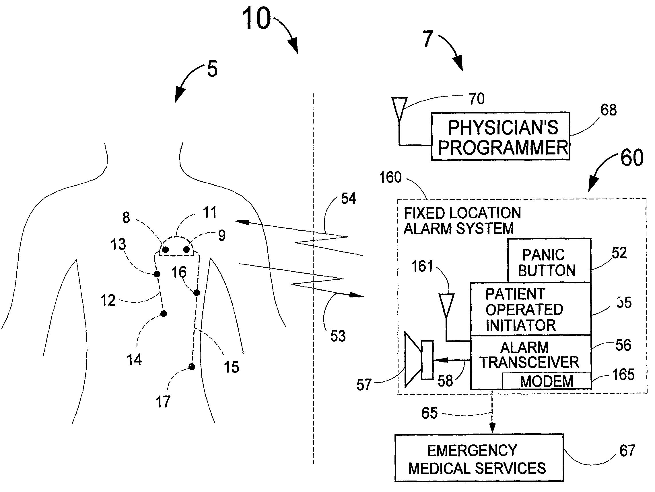
[0079]FIG. 1 illustrates one embodiment of the cardiosaver system 10 consisting of an implanted medical device 5 which is a cardiosaver 5 and external equipment 7. The cardiosaver 5 consists of an electronics module 11 that has two leads 12 and 15 that have multi-wire electrical conductors with surrounding insulation. The lead 12 is shown with two electrodes 13 and 14. The lead 15 has electrodes 16 and 17. In fact, the cardiosaver 5 could utilize as few as one lead or as many as three and each lead could have as few as one electrode or as many as eight electrodes. Furthermore, electrodes 8 and 9 could be placed on the outer surface of the electronics module 11 without any wires being placed externally to the electronics module 11.

[0080]The lead 12 in FIG. 1 could advantageously be placed through the patient's vascular system with the electrode 14 being placed into the apex of the right ventricle. The electrode 13 could be placed in the right ventricle or right atrium or the superior...


PUM


Abstract

Disclosed is a system for detecting an acute myocardial infarction (i.e., a heart attack) at the earliest possible time and promptly warning the patient that he should immediately seek medical care. The present invention includes an implantable electronic system that can sense a change in the patient's electrogram that is indicative of a heart attack. If a heart attack is sensed, the device would then cause an implantable and / or externally located alarm to be actuated to warn the patient of his condition and a medical practitioner at a remote diagnostic center would receive the patient's electrogram for analysis. The patient or a caretaker would then be informed to self-inject medication through a subcutaneous, pass-through drug port that can be a separate device or integrated into the implanted device that is designed for the early detection of a heart attack. Since an implantable heart pacemaker or defibrillator already has within its structure many of the elements required for the device to recognize a heart attack, it would be expeditious to add a capability to these existing devices to detect a heart attack, have a pass-through drug port and provide an implantable and / or external alarm means to inform the patient to take appropriate action.

Description

FIELD OF USE[0001]This invention is in the field of systems, including devices implanted within a human patient, for the purpose of automatically detecting the onset of a cardiac event and promptly providing appropriate medical care to prevent death and / or damage to the heart muscle.BACKGROUND OF THE INVENTION[0002]Heart disease is the leading cause of death in the United States. A common and life-threatening complication of heart disease is myocardial infarction resulting from a thrombus that obstructs blood flow in one or more coronary arteries. The sooner thrombolytic medication such as tissue plasminogen activator (tPA) or urokinase is placed into the patient's bloodstream after the occurrence of an acute myocardial infarction, the sooner an obstructive thrombus will be dissolved and perfusion of the myocardium will be restored. The extent of damage to the myocardium is strongly dependent on the length of time that occurs prior to restoration of some blood flow to the heart musc...

Claims


Application Information

Patent Timeline
Patent Type & Authority Patents(United States)
IPC IPC(8): A61B5/04A61B5/00A61B5/0452A61B5/07A61M25/00A61M39/02A61N1/372
CPCA61B5/0006A61B5/1112A61B5/4839A61B5/6865A61M39/0208A61B5/0031A61B2562/0209A61B5/0452A61B5/076A61M2025/0076A61M2039/0226A61M2205/18A61M2205/3523A61M2205/3584A61M2230/04A61B2505/01A61B2560/0462A61B5/349A61B5/358
Inventor FISCHELL, ROBERT E.FISCHELL, DAVID R.FISCHELL, TIM A.
Owner ANGEL MEDICAL SYST
Learn More