Irreversible electroporation to control bleeding

Inactive Publication Date: 2005-08-04
RGT UNIV OF CALIFORNIA
8 Cites 260 Cited by

AI-Extracted Technical Summary

Problems solved by technology

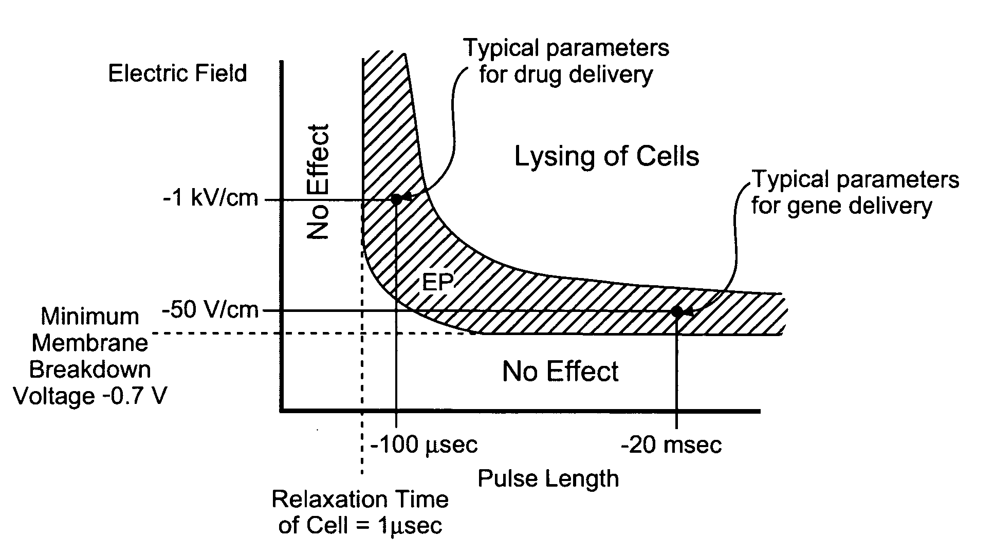
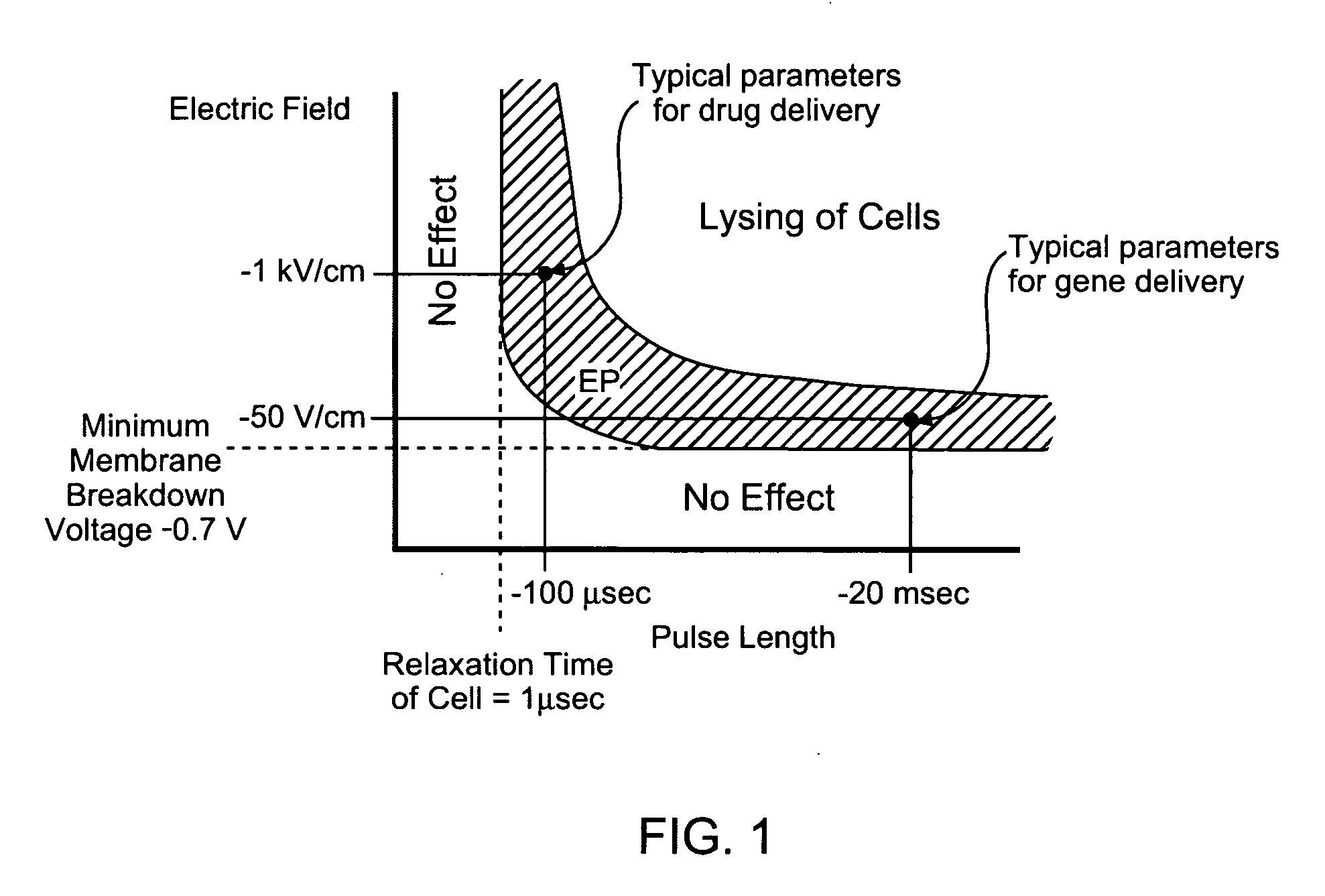
The electroporation pulse should not have considerable thermal effects, but irreversible break cell membranes of targeted cells and result in cell death.
Of these effects, irreversible el...

Benefits of technology

[0020] The present invention comprises a method whereby blood flow and such bleeding from a vessel is stopped with the application of electrical pulses causing irreversible electroporation of cells making up vessels and blood involved in unwanted bleeding. The electric pulses irreversibly permeate the membranes, thereby invoking cell death. The length of time of the electrica...

Abstract

A method of stopping or controlling bleeding by the placement of electrodes into or near the vicinity of vessels is disclosed. Then the application of electrical pulses causing irreversible electroporation of vessel and blood cells throughout the entire area of current flow the bleeding is stopped or controlled. The electric pulses irreversibly permeate the cell membranes, thereby invoking cell death. The irreversibly permeabilized cells are left in situ and are removed by the body immune system. Through the use of irreversible electroporation bleeding can be stopped or controlled without inducing thermal damage.

Application Domain

Surgical needlesInternal electrodes +2

Technology Topic

Cell membraneMedicine +7

Image

  • Irreversible electroporation to control bleeding
  • Irreversible electroporation to control bleeding
  • Irreversible electroporation to control bleeding

Examples

  • Experimental program(3)

Example

Example 1
[0067] The mathematical model provided here shows that irreversible tissue ablation can affect substantial volumes of tissue, without inducing damaging thermal effects. To this end, the present invention uses the Laplace equation to calculate the electrical potential distribution in tissue during typical electroporation pulses and a modified Pennes (bioheat), (Pennes, H. H., Analysis of tissue and arterial blood flow temperatures in the resting forearm. J of Appl. Physiology., 1948. 1: p. 93-122), equation to calculate the resulting temperature distribution. It is important to note that there are several forms of the bioheat equation which have been reviewed (Carney, C. K., Mathematical models of bioheat transfer, in Bioengineering heat transfer, Y. I. Choi, Editor. 1992, Academic Press, Inc: Boston. p. 19-152; Eto, T. K. and B. Rubinsky, Bioheat transfer, in Introduction to bioengineering, S. A. Berger, W. Goldsmith, and E. R. Lewis, Editors. 1996, Oxford Press). While the Pennes equation is controversial, it is nevertheless commonly used because it can provide an estimate of the various biological heat transfer parameters, such as blood flow and metabolism. The modified Pennes equation in this study contains the Joule heating term in tissue as an additional heat source.
[0068] The electrical potential associated with an electroporation pulse is determined by solving the Laplace equation for the potential distribution:
∇·(σ∇φ)=0  (1) [0069] where φ is the electrical potential and σ is the electrical conductivity. The electrical boundary condition of the tissue that is in contact with the leftmost electrode(s) on which the electroporation pulse is applied is:
φ=V0 (2)
[0070] The electrical boundary condition at the interface of the rightmost electrode(s) is:
φ=0  (3)
[0071] The boundaries where the analyzed domain is not in contact with an electrode are treated as electrically insulative to provide an upper limit to the electrical field near the electroporation electrodes and an upper limit to the temperature distribution that results from electroporation: ∂ ϕ ∂ n = 0 ( 4 )
[0072] Solving the Laplace equation enables one to calculate the associated Joule heating, the heat generation rate per unit volume from an electrical field (p):
p=σ|∇φ|2 (5)
[0073] This term is added to the original Pennes equation, (Pennes, H. H., Analysis of tissue and arterial blood flow temperatures in the resting forearm. J of Appl. Physiology., 1948. 1: p. 93-122) to represent the heat generated from the electroporation procedure: ∇ · ( k ⁢ ⁢ Δ ⁢ ⁢ T ) + w b ⁢ ⁢ c b ⁡ ( T a - T ) + q m + p = ρ ⁢ ⁢ c p ⁢ ∂ T ∂ t ( 6 )
[0074] To solve equation (4) it is assumed that the entire tissue is initially at the physiological temperature of 37° C.:
T(x,y,z,0)=37  (7)
[0075] The outer surface of the analyzed domain and the surfaces of the electrodes are taken to be adiabatic, which should produce an upper limit to the calculated temperature distribution in the tissue: ∂ T ∂ n = 0 ⁢ ⁢ ⁢ on ⁢ ⁢ the ⁢ ⁢ electrodes ⁢ ⁢ boundary ⁢ ⁢ and ⁢ ⁢ the ⁢ ⁢ outer ⁢ ⁢ surface ⁢ ⁢ domain ( 8 )
[0076] The analysis modeled conditions typical to tissue electroporation in the liver. The liver was chosen because it is the organ that most minimally invasive ablation techniques treat since cancer in the liver can be resolved by extirpation of the diseased area while surgical resection is not possible in many cases for this organ (Onik, G., B. Rubinsky, and et al., Ultrasound-Guided Hepatic Cryosurgery in the Treatment of Metastatic Colon Carcinoma. Cancer, 1991. 67(4): p. 901-907). The electroporation parameters, i.e. pulse parameters for reversible and irreversible electroporation where obtained from rat liver data (Miklavcic, D., et al., A validated model of in vivo electric field distribution in tissues for electrochemotherapy and for DNA electrotransfer for gene therapy. Biochimica et Biophysica Acta, 2000. 1523(1): p. 73-83; Suzuki, T., et al., Direct gene transfer into rat liver cells by in vivo electroporation. FEBS Letters, 1998. 425(3): p. 436-440), but biological parameters corresponding to the human liver were used in the analysis. Tissue thermal properties are taken from reference (Duck, F. A., Physical Properties of Tissues: A Comprehensive Reference Book. 1990, San Diego: Academic Press) and the electrical properties from reference (Boone, K., D. Barber, and B. Brown, Review—Imaging with electricity: report of the European Concerted Action on Impedance Tomography. J. Med. Eng. Technol., 1997.21: p. 201-232) and are listed in table 1. The tissue is assumed isotropic and macroscopically homogeneous.The intent of the analysis was to determine the extent of the region in which reversible or irreversible electroporation is induced in the liver for various electroporation voltages and durations while the maximal temperature in the tissue is below 50° C. Thermal damage is a time-dependent process described by an Arhenius type equation (Henriques, F. C. and A. R. Moritz, Studies in thermal injuries: the predictability and the significance of thermally induced rate processes leading to irreversible epidermal damage. Arch Pathol., 1947. 43: p. 489-502; Diller, K. R., Modeling of bioheat transfer processes at high and low temperatures, in Bioengineering heat transfer, Y. I. Choi, Editor. 1992, Academic Press, Inc: Boston. p. 157-357),
Ω=∫ξe−E a /RT dt (9)
[0077] Where Ω is a measure of thermal damage, ξ is the frequency factor, Ea is the activation energy and R is the universal gas constant. A detailed description on the various degrees of thermal damage as described in Equation (9) above can be found in (Diller, K. R., Modeling of bioheat transfer processes at high and low temperatures, in Bioengineering heat transfer, Y. I. Choi, Editor. 1992, Academic Press, Inc: Boston. p. 157-357).
[0078] A careful examination shows that the thermal damage is a complex function of time, temperature and all the parameters in Equation (9) above and that there are various degrees of thermal damage. In various applications or for various considerations it is possible to design irreversible electroporation protocols that induce some degree of thermal damage, either in part of the electroporated region or at a reduced level throughout the electroporated region. However, in this example we have chosen 50° C. as the target temperature for several reasons. Thermal damage begins at temperatures higher than 42° C., but only for prolonged exposures. Damage is relatively low until 50° C. to 60° C. at which the rate of damage dramatically increases (Diller, K. R., Modeling of bioheat transfer processes at high and low temperatures, in Bioengineering heat transfer, Y. I. Choi, Editor. 1992, Academic Press, Inc: Boston. p. 157-357). Therefore 50 C will be a relatively low bound on the possible thermal effects during irreversible electroporation. It is anticipated that the electrical parameters chosen for irreversible electroporation without a thermal effect could be substantially longer and higher than those obtained from an evaluation for 50 C in this example. Furthermore, since the Laplace and bioheat equations are linear, the results provided here can be extrapolated and considered indicative of the overall thermal behavior.
[0079] The analyzed configurations have two needles or four needle electrodes embedded in a square model of the liver. Needle electrodes are commonly used in tissue electroporation and will be most likely also used in the liver (Somiari, S., et al., Theory and in vivo application of electroporative gene delivery. Molecular Therapy, 2000. 2(3): p. 178-187). The square model of the liver was chosen large enough to avoid outer surface boundary effects and to produce an upper limit for the temperature, which develops during electroporation in the liver. For each configuration the surface of one electrode is assumed to have a prescribed voltage with the other electrode set to ground. The effect of the spacing between the electrodes was investigated by comparing distances of 5, 7.5 and 10 mm, which are typical. The electrodes were also modeled with typical dimensions of 0.5, 1 and 1.5 mm in diameter. The blood flow perfusion rate was taken to zero or 1.0 kg/M3 s (Deng, Z. S. and J. Liu, Blood perfusion-based model for characterizing the temperature fluctuations in living tissue. Phys A STAT Mech Appl, 2001. 300: p. 521-530). The metabolic heat was taken to be either zero or 33.8 kW/m3 (Deng, Z. S. and J. Liu, Blood perfusion-based model for characterizing the temperature fluctuations in living tissue. Phys A STAT Mech Appl, 2001. 300: p. 521-530).
[0080] The calculations were made for an electroporation pulse of 800 μs. This pulse duration was chosen because typically, reversible electroporation is done with eight separate 100 μs pulses, (Miklavcic, D., et al., A validated model of in vivo electric field distribution in tissues for electrochemotherapy and for DNA electrotransfer for gene therapy. Biochimica et Biophysica Acta, 2000. 1523(1): p. 73-83) and therefore the value we chose is an upper limit of the thermal effect in a pulse time frame comparable to that of reversible electroporation. Consequently, the results obtained here are the lower limit in possible lesion size during irreversible electroporation. It should be emphasized that we believe irreversible electroporation tissue ablation can be done with shorter pulses than 800 μs. To evaluate the thermal effect, we gradually increased in our mathematical model the applied pulse amplitude for the 800 μs pulse length until our calculations indicated that the electroporation probe temperature reached 50° C., which we considered to be the thermal damage limit. Then, we evaluated the electric field distribution throughout the liver.
[0081] A transmembrane potential on the order of 1V is required to induce irreversible electroporation. This value is dependent on a variety of conditions such as tissue type, cell size and other external conditions and pulse parameters. The primary electrical parameter affecting the transmembrane potential for a specific tissue type is the amplitude of the electric field to which the tissue is exposed. The electric field thresholds used in estimating the extent of the region that was irreversibly electroporated were taken from the fundamental studies of Miklavcic, Mir and their colleagues performed with rabbit liver tissue (Miklavcic, D., et al., A validated model of in vivo electric field distribution in tissues for electrochemotherapy and for DNA electrotransfer for gene therapy. Biochimica et Biophysica Acta, 2000. 1523(1): p. 73-83). In this study, that correlated electroporation experiments with mathematical modeling, they have found that the electric field for reversible electroporation is 362 +/−21 V/cm and is 637 +/−43 V/cm for irreversible electroporation for rat liver tissue. Therefore, in the analysis an electric field of 360 V/cm is taken to represent the delineation between no electroporation and reversible electroporation and 680 V/cm to represent the delineation between reversible and irreversible electroporation.
[0082] All calculations were performed using MATLAB's finite element solver, Femlab v2.2 (The MathWorks, Inc. Natick, Mass.). To ensure mesh quality and validity of solution, the mesh was refined until there was less than a 0.5% difference in solution between refinements. The baseline mesh with two 1 mm electrodes, 10 mm spacing had 4035 nodes and 7856 triangles. The simulations were conducted on a Dell Optiplex GX240 with 512 MB of RAM operating on Microsoft Windows 2000.
Results and Discussion
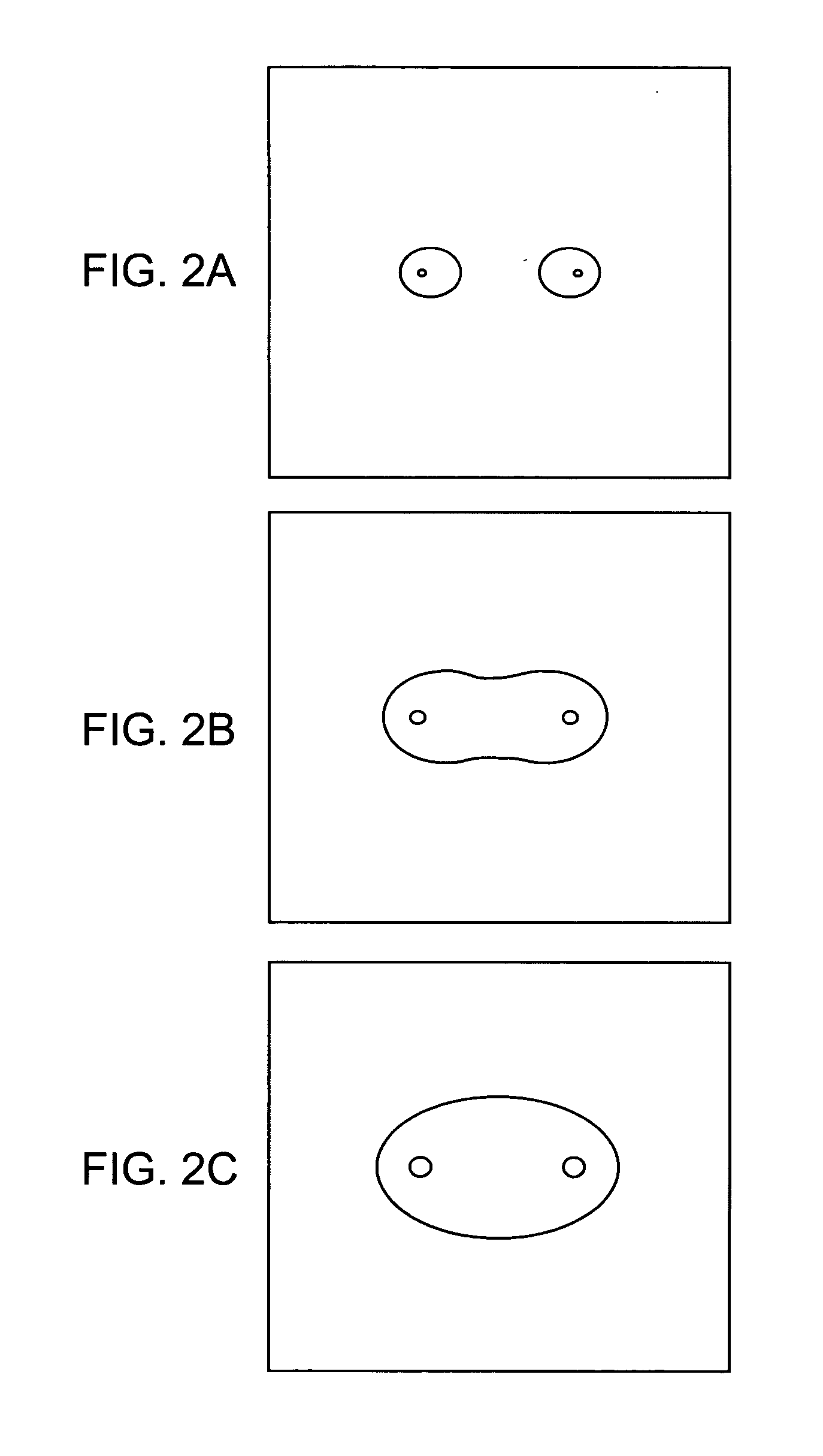
[0083]FIGS. 2 and 3 examine the effect of the electrode size and spacing on the ablated area in a two-needle electroporation configuration. In obtaining these figures, we ignored the effect of the blood flow and metabolism in the heat transfer equation, which should give an upper limit for the estimated ablation area. FIG. 2 compares the extent of the irreversible electroporated area for electroporation electrode sizes of 0.5, 1 and 1.5 mm in diameter and a distance between electrodes of 10 mm. The strong effect of the electrode size is evident. It is seen that for the smaller electrodes, the irreversibly electroporated area is not contiguous, while for a 1.5 mm electrode the area of potential tissue, ablation has an elliptical shape with dimensions of about 15 mm by 10 mm. In the brackets, we give the electroporation voltage for which the probe temperature reaches 50° C. in these three configurations. It is seen that the range is from 857V for the 0.5 mm probe to 1575V for the 1.5 mm probe. This is within the typical range of tissue electroporation pulses. FIG. 3 evaluates the effect of the spacing between the electrodes. It is observed that in the tested range, the small dimension of the contiguous elliptical shape of the ablated lesion remains the same, while the larger dimension seems to scale with the distance between the electrodes.
[0084]FIGS. 2 and 3 demonstrate that the extent of tissue ablation with irreversible electroporation is comparable to that of other typical minimally invasive methods for tissue ablation, such as cryosurgery (Onik, G. M., B. Rubinsky, and et. al., Ultrasound-guided hepatic cryosurgery in the treatment of metastatic colon carcinoma. Cancer, 1991. 67(4): p. 901-907; Onik, G. M., et al., Transrectal ultrasound-guided percutaneous radical cryosurgical ablation of the prostate. Cancer, 1993. 72(4): p. 1291-99). It also shows that varying electrode size and spacing can control lesion size and shape. The shape and size of the ablated lesion can be also controlled by varying the number of electrodes used. This is shown in FIGS. 4 and 5, for a four-electrode configuration. These figures also compare the effect of probe size and spacing and the results were also obtained by ignoring the effect of blood flow and metabolism in the energy equation. Again, it is seen that larger electrodes have a substantial effect on the extent of the ablated region and that the extent of ablation scales with the spacing between the electrodes.
[0085] A comparison between reversible and irreversible electroporation protocols can be achieved from FIGS. 6 and 7. In FIG. 6, an 800 μs, 1295 V pulse was applied between two 1.5 mm diameter electrodes placed 10 mm apart. This produces a tissue temperature lower than 50° C. The figure plots the margin of the irreversibly electroporated region, i.e. the 680 V/cm voltage-to-distance gradients and that of the reversible electroporated region, the 360 V/cm gradients. FIG. 7 was obtained for two 1 mm electrodes placed 10 mm apart. In this figure, we produced an electroporated region that was only reversibly electroporated, i.e. with electric fields lower than 360 V/cm. In comparing FIGS. 6 and 7, it is obvious that the extent of the ablated area possible through electrochemotherapy alone is substantially smaller than that through irreversible electroporation alone.
[0086] The effect of blood flow and metabolism on the extent of irreversible electroporation is illustrated in FIG. 8. The figures compare a situation with metabolism and a relatively high blood flow rate to a situation without blood flow or metabolism. It is obvious that metabolism and blood perfusion have a negligible effect on the possible extent of irreversible tissue electroporation. This is because the effect of the Joule heating produced by the electroporation current is substantially larger than the effects of blood flow or metabolism.
[0087] An even more conservative estimate for the thermal damage can be obtained by assuming that the tissue reaches 50° C. instantaneously, during the electroporation pulses such that the damage is defined as
Ω=tpξe−ΔE/RT (10)
[0088] Several values taken from the literature for activation energy and frequency factor were applied to equation (10) with the pulse lengths calculated in the examples above. Because the application of the pulse is so short, the damage would be near zero, many times less than the value (Ω=0.53) to induce a first degree burn (Diller, K. R., Modeling of bioheat transfer processes at high and low temperatures, in Bioengineering heat transfer, Y. I. Choi, Editor. 1992, Academic Press, Inc: Boston. p. 157-357). regardless of the values used for activation energy and frequency factor.
[0089] Currently, tissue ablation by electroporation is produced through the use of cytotoxic drugs injected in tissue combined with reversible electroporation, a procedure known as electrochemotherapy. The present invention shows that irreversible electroporation by itself produces substantial tissue ablation for the destruction of undesirable tissues in the body. The concern was that higher voltages required for irreversible electroporation would cause Joule heating and would induce thermal tissue damage to a degree that would make irreversible electroporation a marginal effect in tissue ablation. Using a mathematical model for calculating the electrical potential and temperature field in tissue during electroporation, the present invention shows that the area ablated by irreversible tissue electroporation prior to the onset of thermal effects is substantial and comparable to that of other tissue ablation techniques such as cryosurgery. Our earlier studies have shown that the extent of electroporation can be imaged in real time with electrical impedance tomography (Davalos, R. V., B. Rubinsky, and D. M. Otten, A feasibility study for electrical impedance tomography as a means to monitor tissue electroporation for molecular medicine. IEEE Transactions on Biomedical Engineering, 2002. 49(4): p. 400-403; Davalos, R. V., et al., Electrical impedance tomography for imaging tissue electroporation. IEEE Transactions on Biomedical Engineering, 2004). Irreversible electroporation, therefore, has the advantage of being a tissue ablation technique, which is as easy to apply as high temperature ablation, without the need for adjuvant chemicals as required in electrochemical ablation and electrochemotherapy. In addition, a unique aspect of irreversible electroporation is that the affected area can be controlled in real time with electrical impedance tomography.

Example

Example 2
[0090] This example was developed to produce a correlation between electroporation pulses and thermal effects. The system analyzed is an infinitesimally small control volume of tissue exposed to an electroporation voltage gradient of V (Volts/cm).The entire electrical energy is dissipated as heat and there is no conduction of heat from the system. The calculations produce the increase in temperature with time during the application of the pulse and the results are a safe lower limit for how long a certain electroporation pulse can be administered until a certain temperature is reached. To generate the correlation an energy balance is made on a control volume between the Joule heating produced from the dissipation of heat of the V (volt/cm) electrical potential dissipating through tissue with an electrical conductivity of σ ( ohm-cm) and the raise in temperature of the control volume made of tissue with a density ρ (g/cc) and specific heat, c, (J/g K). the calculation produces the following equation for the raise in temperature (T) per unit time (t) as a function of the voltage gradients and the thermal and electrical properties of the liver. ⅆ T ⅆ t = V 2 ⁢ σ ρ ⁢ ⁢ c ( 2 ⁢ - ⁢ 1 )
[0091] The table below was obtained for the liver with the following properties: [0092] Electrical resistivity of liver—8.33 Ohm-meter [0093] Specific heat of liver—J/g K [0094] Density of liver—1 g/cc
[0095] We obtain the following table: TABLE 1 Voltage Time per degree time from 37 C. to Gradient - V (V/cm) C. rise (ms) 65 C. (ms) 50 1199.52 33586.56 100 299.88 8396.64 150 133.28 3731.84 200 74.97 2099.16 250 47.98 1343.46 300 33.32 932.96 350 24.48 685.44 400 18.74 524.79 450 14.81 414.65 500 12.00 335.87 550 9.91 277.57 600 8.33 233.24 650 7.10 198.74 700 6.12 171.36 750 5.33 149.27 800 4.69 131.20 850 4.15 116.22 900 3.70 103.66 950 3.32 93.04 1000 3.00 83.97 1050 2.72 76.16 1100 2.48 69.39 1150 2.27 63.49 1200 2.08 58.31 1250 1.92 53.74 1300 1.77 49.68 1350 1.65 46.07 1400 1.53 42.84 1450 1.43 39.94 1500 1.33 37.32
[0096] The second column of Table 1 gives the amount of time it takes for the temperature of the liver to raise 1 C, when the tissue experiences the electroporation pulse in column 1. The time for even a relatively high electroporation voltage of 1500V/cm is of the order of 1.33 millisecond for 1 C rise and 37.32 millisecond until a temperature of 65 C is reached. Using the equation (2-1) or Table 1 it is possible to evaluate the amount of time a certain pulse can be applied without inducing thermal effects. Considering the typical electroporation parameters reported so far there is no limitation in the electroporation length from thermal considerations. Column 3 of Table 1 shows the time required to reach 65 C, which is where thermal damage may begin. The calculations in this example give a lower limit for the extent of time in which a certain thermal effects will be induced by electroporation pulses. For more precise calculations it is possible to use the equation developed in this example with equation (9) or (10) from Example 1.

Example

Example 3
[0097] The goal of this experiment was to verify the ability of irreversible electroporation pulses to produce substantial tissue ablation in the non-thermal regime. To this end we have performed experiments on the liver of Spraque-Dawley male rats (250 g to 350 g) under an approved animal use and care protocol. After the animals were anesthetized by injection of Nembutal Sodium Solution (50 mg/ml Pentobarbital) the liver was exposed via a midline incisions and one lobed clamped between two cylindrical electrodes of Ag/AgCl, with a diameter of 10 mm (In Vivo Metric, Healdsburg, Calif.). The electrodes had their flat surface parallel; they were concentric and the liver between the electrodes was compressed so that the lobes were separated by 4 mm. A schematic of the electrodes and the liver is shown in FIG. 9. The liver was exposed to a single electroporation pulse of 40 milliseconds. One electrode was set to 400 V and the other grounded. The rest of the liver was not in contact with any media and therefore is considered electrically insulated. After electroporation the rat was maintained under controlled anesthesia for three hours. Following exsanguination the liver was flushed with physiological saline under pressure and fixed by perfusion with formaldehyde. The liver was resected through the center of the electroporated region and analyzed by histology. FIGS. 10 and 11 show the appearance of the liver. Histology has determined that the dark area corresponds to the region of tissue necrosis. The electrical field in the electroporated liver and the temperature distribution were calculated using the equations in Example 1, subject to one electrode at a voltage of 400V and the other grounded, for 40 milliseconds. The liver was modeled as an infinite slab of 4 mm thickness, with concentric cylindrical electrodes (see FIG. 9). The results are shown in FIG. 12. FIG. 12 shows lines of constant voltage gradients (V/cm) and lines of constant temperature. It is evident that in the majority of the electroporated tissue the temperature is about 42 C immediately after the pulse. The highest temperature occurs near the edge of the cylindrical electrodes, where it is about 50 C. FIG. 13 was obtained by bringing together FIGS. 11 and 12. Superimposing the calculated results on the histological measurements reveals that the dark (necrotic) area margin corresponds to electroporation parameters of about 300 V/cm. The results demonstrate that irreversible electroporation can induce substantial tissue necrosis without the need for chemical additives as in electrochemotherapy and without a thermal effect.
[0098] The results obtained by disrupting blood flow to a given area are dramatically shown in FIG. 14 which is a photo of a micrograph. This micrograph is from the interface between irreversible electroporated liver and normal liver. The left hand side shows normal hepatocytes with clear nucleus and nuclei. The photo shows well defined cell membranes and clean (flushed) sinusoids. The right hand side of FIG. 14 shows condensed nuclei, no evidence of cell membrane, expanded cell border with no evidence of sinusoids. The disintegrated red blood cells shown in FIG. 14 are in what could have been the spaces of the sinusoids. Flushing is not believed to have had an effect on the results obtained on the right hand side of FIG. 14.
[0099] The preceding merely illustrates the principles of the invention. It will be appreciated that those skilled in the art will be able to devise various arrangements which, although not explicitly described or shown herein, embody the principles of the invention and are included within its spirit and scope. Furthermore, all examples and conditional language recited herein are principally intended to aid the reader in understanding the principles of the invention and the concepts contributed by the inventors to furthering the art, and are to be construed as being without limitation to such specifically recited examples and conditions. Moreover, all statements herein reciting principles, aspects, and embodiments of the invention as well as specific examples thereof, are intended to encompass both structural and functional equivalents thereof. Additionally, it is intended that such equivalents include both currently known equivalents and equivalents developed in the future, i.e., any elements developed that perform the same function, regardless of structure. The scope of the present invention, therefore, is not intended to be limited to the exemplary embodiments shown and described herein. Rather, the scope and spirit of present invention is embodied by the appended claims.

PUM

no PUM

Description & Claims & Application Information

We can also present the details of the Description, Claims and Application information to help users get a comprehensive understanding of the technical details of the patent, such as background art, summary of invention, brief description of drawings, description of embodiments, and other original content. On the other hand, users can also determine the specific scope of protection of the technology through the list of claims; as well as understand the changes in the life cycle of the technology with the presentation of the patent timeline. Login to view more.
Why Eureka
Try Eureka