Automatic x-ray detection for intra-oral dental x-ray imaging apparatus

a dental x-ray imaging and automatic detection technology, applied in the field of automatic x-ray detection for intraoral dental x-ray imaging apparatus, can solve the problems of inherently more expensive arrangement, negative impact on diagnostic image noise performance, and lack of collateral advantage of removing accumulated dark curren

Inactive Publication Date: 2006-03-21
DENTAL IMAGING TECH
View PDF134 Cites 21 Cited by
  • Summary
  • Abstract
  • Description
  • Claims
  • Application Information

AI Technical Summary

Benefits of technology

[0011]The object of the invention is an automatic x-ray detection apparatus and method for x-ray digital imagers for dental and medical application, capable of producing automatic triggering of the imager to the integration mode with immediate reaction to the start of the x-ray emission, and including provision against occurrence of false triggering caused by variations in the image sensor output signal due to abnormal imager defects and / or variations of the ambient and temperature conditions.
[0012]The method adopted will ensure at the same time effective removal of the dark current spontaneously generated in the imager, with minimised impact on the power consumption of the imaging system, while the apparatus will perform its function without need of electrical connections to the x-ray generator and will use the processing power of the existing microcontroller, without need of additional comparator circuitry.

Problems solved by technology

For the intrinsic characteristics of the imager, usually a remarkable dark current is spontaneously generated, which is having a negative impact on the noise performance of the diagnostic image.
Such arrangement is clearly disadvantageous for the need of having an electrical connection with the x-ray generator.
Such arrangement is inherently more expensive, although characterised by a prompt reaction time, and does not provide the collateral advantage of removing the accumulated dark current as provided by the continuous clocking and readout method.
This arrangement is disadvantageous because it requires an external analogue circuit for the comparison and does not allow for sophisticated processing of the video output in order to prevent false triggering caused by abnormal imager defects.

Method used


Image

  • Automatic x-ray detection for intra-oral dental x-ray imaging apparatus
  • Automatic x-ray detection for intra-oral dental x-ray imaging apparatus
  • Automatic x-ray detection for intra-oral dental x-ray imaging apparatus

Examples


Embodiment Construction

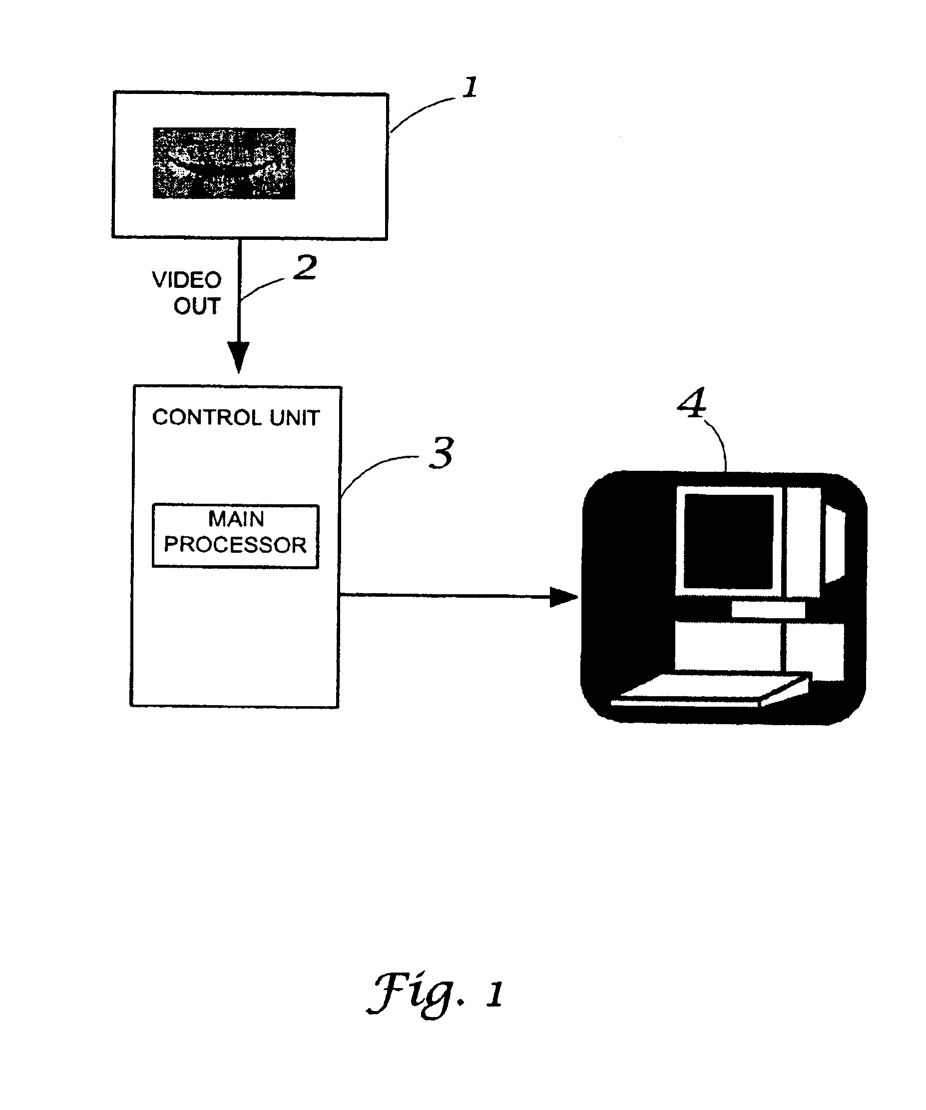
[0017]The system illustrated in FIG. 1 is a typical x-ray diagnostic system dedicated to dental intra-oral radiography with digital image acquisition.

[0018]The x-ray source 1 is aligned with the image receptor 2 (the x-ray imager) by means of a suitable alignment device. The imager is located in the patient mouth, behind the object (tooth) to be imaged. It is connected to the control unit 3, providing imager control and image acquisition and transfer to the main processor 4 (i.e. the Personal Computer), where the diagnostic image display, processing and archive is performed.

[0019]The x-ray imager is a solid state device including a matrix of radiation sensitive pixels.

[0020]Solid state imagers, such as CCD devices, are in general exhibiting spontaneous generation of dark current, which is in turn generating a noise signal increasing, by know relations, with the temperature and with the time.

[0021]As this noise signal may significantly use the signal range of the device, it is impera...


PUM


Abstract

An apparatus and method used in intra-oral dental x-ray imaging equipment provides automatic detection of the x-ray emission in order to obtain timely transition to the image integration and acquisition phase with high level of rejection of false triggering induced by blemish defects of the image sensor or by variations of the ambient and temperature conditions. A solid state imager, such as a CCD image sensor, is continually clocked during the standby phase prior to irradiation from an X-ray source, so providing on same time an output signal proportional to the accumulated dark current and the continuous removal of the same. A control unit analyses the output signal of the imager, detects the variation caused by the start of the x-ray emission using appropriate threshold levels, and automatically triggers the imager to the image integration and acquisition phases. Based on memorised maps of the imager blemish defects, and continually monitored variations of the imager output signal, the control unit will reject false triggering due to imager defects or variations of the ambient and temperature conditions.

Description

[0001]This application is a continuation of application Ser. No. 09 / 754,663, filed Jan. 4, 2001, U.S. Pat. No. 6,775,351, which claims the benefit of U.S. Provisional Application 60 / 179,639, filed Feb. 2, 2000.BACKGROUND OF THE INVENTION[0002]CCD and other types of solid state imagers have got widespread use in dental and medical radiology for their ability to provide in real time x-ray images of high diagnostic value, with reduced levels of the X-ray dose imparted to the patient versus the conventional radiographic film.[0003]In the most typical arrangement the imager is in a wait state, and requires to be triggered into an integration state as soon as the irradiation starts, where the imager clocking is suspended and the x-ray conversion charge is accumulated.[0004]It is one desirable feature that the triggering to the integration state occurs in an automatic way, without need of connections to the x-ray generator, using as low as possible x-ray dose threshold to minimise useless ...

Claims


Application Information

Patent Timeline
Patent Type & Authority Patents(United States)
IPC IPC(8): H05G1/64A61B6/14H04N5/32H04N5/361H04N5/367
CPCA61B6/145A61B6/4233H04N5/367H04N5/361H04N5/32H04N25/63H04N25/68
Inventor RINALDI, GERARDOGIANFRANCO, VENTURINOROTONDO, GIUSEPPE
Owner DENTAL IMAGING TECH
Why Eureka
Try Eureka