Bonding of soft biological tissues by passing high frequency electric current therethrough

a technology of high frequency electric current and soft biological tissue, which is applied in the field of bonding soft biological tissue with high frequency electric current, can solve the problems of delayed healing and/or inflammation, allergic reactions, limited applicability, etc., and achieve the effect of accurately controlling the degree of coagulation and promoting healing

Inactive Publication Date: 2006-04-11
BIOFUSE MEDICAL TECH +1
View PDF47 Cites 125 Cited by
  • Summary
  • Abstract
  • Description
  • Claims
  • Application Information

AI Technical Summary

Benefits of technology

[0011]Another object of the invention is to prevent sticking of the electrodes to the tissue.
[0013]Yet another object of the invention is to prevent burning of tissue in the bipolar electrode zone.
[0015]Still another object of the invention is to bond tissue to close an incision quickly and reliably.
[0016]Another object of the invention is to bond tissue in a way which promotes fast healing.
[0017]A further object of the invention is to rely on measurement of tissue impedance to accurately control the degree of coagulation which bonds the tissue for a wide variety of different tissues.
[0022]Another aspect of the present invention is directed to a method and apparatus for bonding soft biological tissue having an incision therein with forceps adapted to grip a portion of the tissue on both sides of the incision. Electrodes are provided for contacting the tissue portion. An electrical power source provides a high frequency electrical signal to the electrodes to be passed through the tissue portion. The electrodes are dimensioned relative to size of the tissue portion to be an effective heat sink for conducting heat away from the tissue and thereby prevent sticking of tissue to the electrodes.

Problems solved by technology

These techniques have a number of well known disadvantages which include one or more of the following: leaving a foreign body in the tissue, pinching of tissue which can cause delayed healing and / or inflammation, allergic reaction, limited applicability, complexity of use, and the need for expensive equipment.
However, at temperature exceeding 100° C. the tissue becomes dehydrated, its electric resistance increases, which leads to further temperature rise and charring of the tissue.
Still this technique has not been accepted for general clinical use because of the technical complexity of its utilization and because of inadequate surface energy release.
However, the use of the existing bipolar devices for connecting soft tissues other than walls of compressed blood vessels encounters insurmountable difficulties.
Specifically, it has been difficult to correctly set the electrical signal parameters to achieve such aims.
If too little current is applied, then the tissue joint can be spongy, weak and unreliable.
On the other hand, if too much current is applied, then the working surface of the electrode can stick to the tissue so that removal of the electrode causes bleeding and possible injury.
Also, the tissue in the overly-heated zone can become desiccated and charred.
Therefore, such high frequency coagulative devices have seen limited use for only hemostasis of blood vessels of relatively small diameter.
Unless the heating is stopped, severe tissue damage will occur.
Usage of these techniques for other types of tissue creates severe difficulties due to the wide variation in values of impedance which can be encountered due to, for example, tissue structure, thickness, condition of the tissue and condition of the tool surface.

Method used


Image

  • Bonding of soft biological tissues by passing high frequency electric current therethrough
  • Bonding of soft biological tissues by passing high frequency electric current therethrough
  • Bonding of soft biological tissues by passing high frequency electric current therethrough

Examples


Embodiment Construction

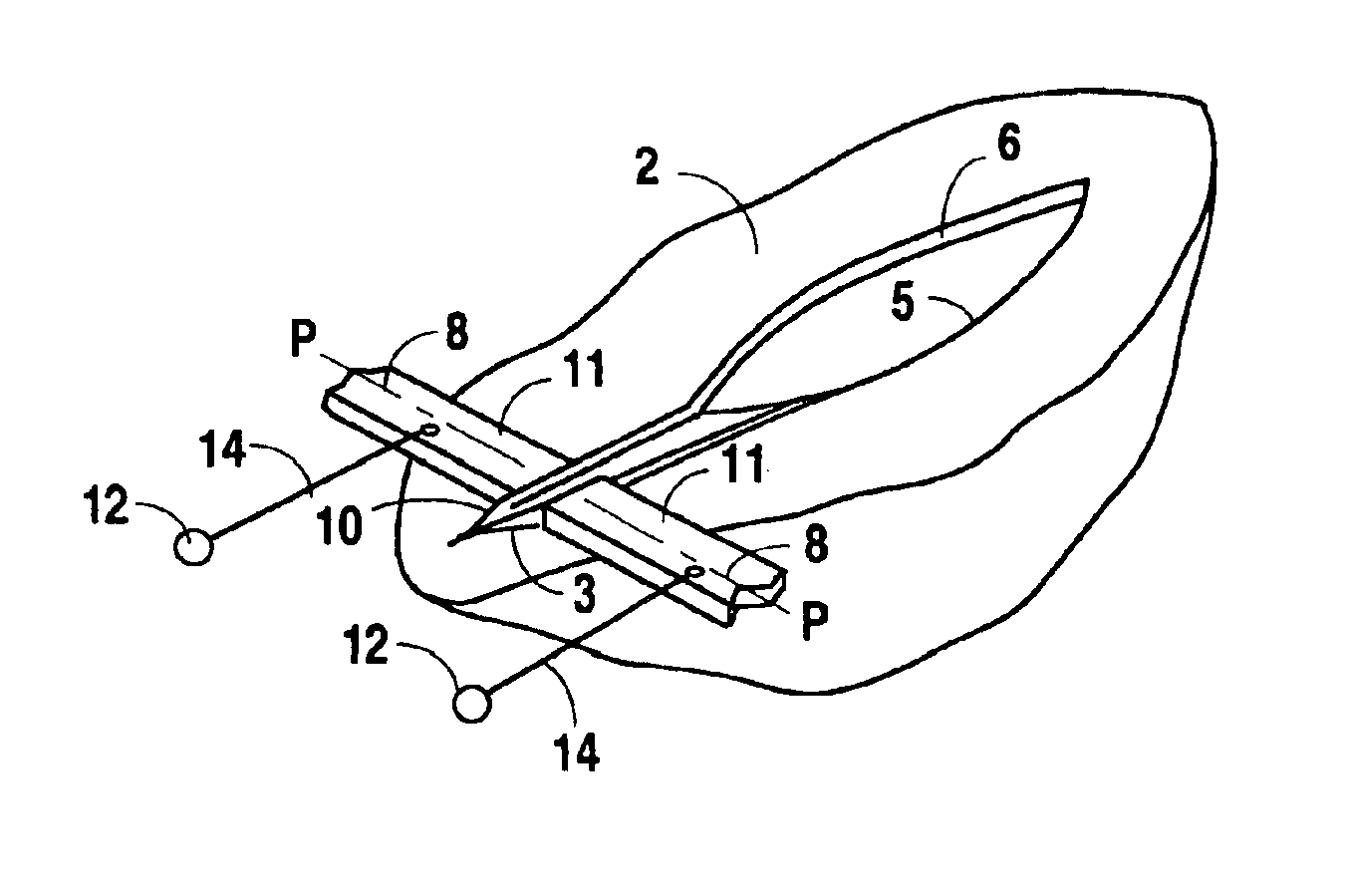
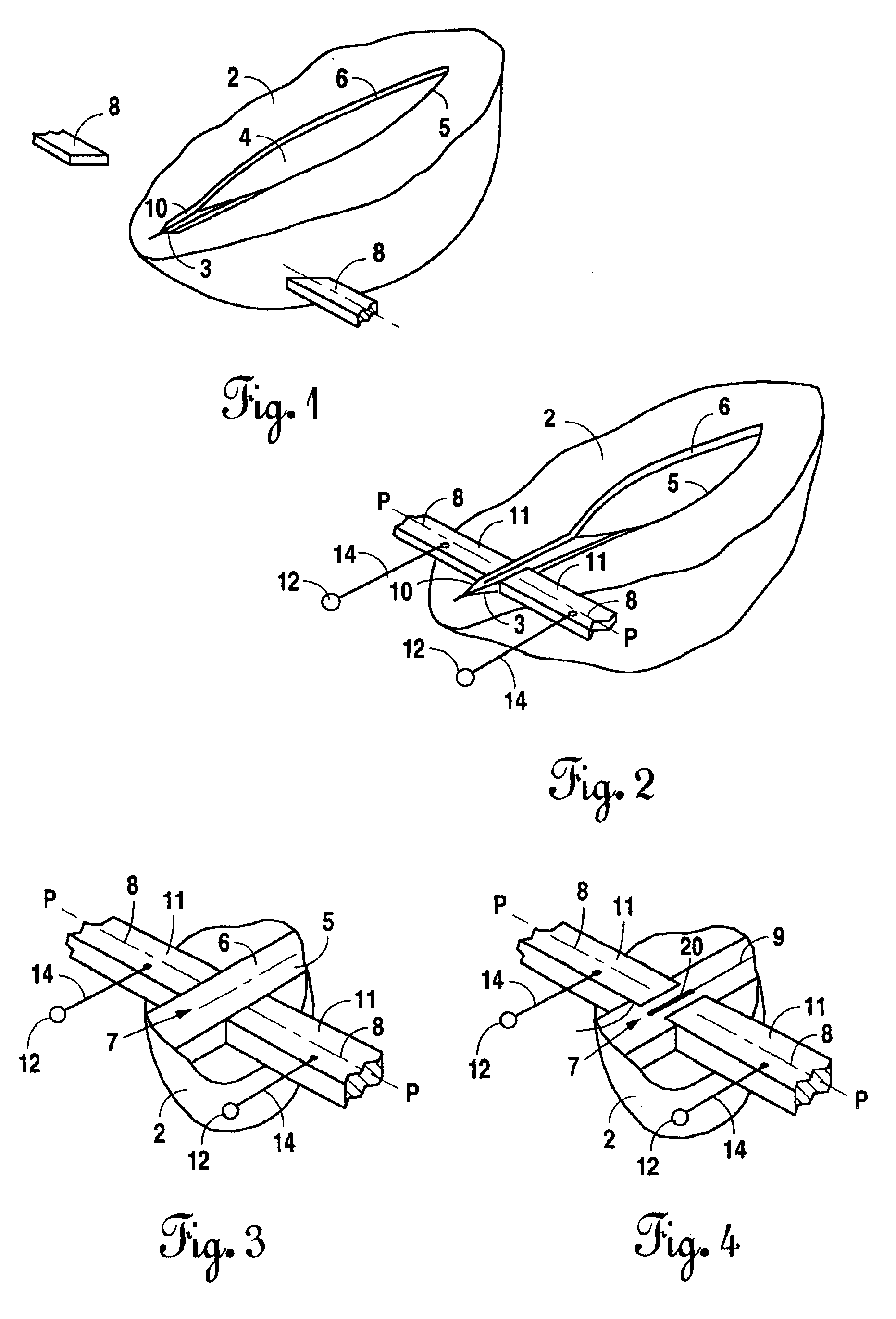
[0043]FIG. 1 shows tissue 2 with an incision 4 formed therein. Incision 4 could have been formed as part of some surgery done on a patient, or it could be an injury due to some type of trauma. The incision can be a cut in the skin or in a wall of an organ, or the organ itself, e.g. a blood vessel or nerve. In any case, the incision must be closed by bonding, or joining, the edges of tissue 5 and 6 on either side of the incision to each other.

[0044]In accordance with the present invention, the edges 5, 6 at end 3 of the incision are gripped and raised by pincers (not shown) to form tissue portion 10 in the form of a flange. This is depicted in FIG. 1. A forceps tool (referred to herein as a forceps) is provided in the form of any instrument capable of gripping the tissue and selectively adding a clamping force under manual control. Various forceps designs are well known. Typically they include a pair of arms with opposed ends between which the tissue can be gripped. Forceps arranged ...


PUM


Abstract

Technique for bonding soft biological tissue having an incision therein with forceps adapted to grip a portion of tissue on both sides of incision. Electrodes are secured to forceps for contracting the tissue portion. An electrical power source provides a high frequency electrical signal to electrodes to be passed through the tissue portion. The electrical power source is controlled to provide electrodes with one voltage signal during a first of two stages, wherein the voltage rises linearly, and another voltage signal during a second of the two stages, wherein the voltage is stabilized and modulated with a low frequency rectangular signal. A clamping means applies force with the forceps to compress the tissue at one or different levels during two time periods while the high frequency voltage is passed through the electrodes. The tissue impedance is measured as a function of time, with its minimal value being determined and stored.

Description

[0001]This application is a Divisional of Ser. No. 09 / 022,869 filed Feb. 12, 1998 now U.S. Pat. No. 6,562,037.BACKGROUND OF THE INVENTION[0002]The present invention is directed to a technique for bonding soft biological tissue to close an incision therein and, in particular, to heating of the tissue with high frequency electric current in combination with compression of the tissue.[0003]For purposes of the ensuing discussion, soft biological tissue will be referred to just by the term “tissue” for reasons of simplicity and economy of space, and should be understood to mean any tissue other than bone, such as skin, organs, blood vessels and nerves. When tissue is injured, it must be repaired by re-joining the edges of tissue that has been torn or cut. For example, when tissue is cut during a surgical operation, the incision(s) must be closed to complete the surgery. In fact, a tissue break (particularly in blood vessels) may also need to be closed even during surgery, such as to prov...

Claims


Application Information

Patent Timeline
Patent Type & Authority Patents(United States)
IPC IPC(8): A61B18/18A61B18/12A61B18/14
CPCA61B18/1442A61B2018/00619A61B2018/0063A61B2019/304A61B2018/00672A61B2018/00678A61B2018/1462A61B2018/00666A61B2090/034
Inventor PATON, BORIS E.LEBEDEV, VLADIMIR K.VORONA, DAVID S.KARCHEMSKY, VOLODIMIR I.FURMANOV, YURI A.LEBEDEV, ALEXSEY V.VASILCHENKO, VALERY A.SIDORENKO, DMITRY F.IEMCHENKO-RIBKO, VITALY P.IVANOVA, OLGA N.FURMANOV, ALEXANDR Y.ZHYVODERNIKOV, YEVGEN V.LYASHENKO, ANDREI A.SAVITSKAYA, IRINA M.
Owner BIOFUSE MEDICAL TECH
Why Eureka
Try Eureka