Multi energy x-ray imager

a multi-energy, x-ray technology, applied in the field of image acquisition, can solve the problems of increasing the idle time of the apparatus, affecting the efficiency of the facility, increasing the operating and overhead costs of the medical diagnostic facility, etc., and achieves the effect of high energy level and efficient collection of charges

Inactive Publication Date: 2006-05-30
VARIAN MEDICAL SYSTEMS
View PDF7 Cites 17 Cited by
  • Summary
  • Abstract
  • Description
  • Claims
  • Application Information

AI Technical Summary

Benefits of technology

[0009]In accordance with an embodiment of the present invention, the conversion panel includes a first electrode, a second electrode, and a photoconductor secured between the first and second electrodes. By way of non-limiting examples, the photoconductor can be made from Mercuric Iodide (HgI2), Lead Iodide (PbI2), Bismuth Iodide (BiI3), Cesium Iodide (CsI), Cadmium Zinc Telluride (CdZnTe), or equivalents thereof. The photoconductor preferably has a thickness configured for absorbing X-ray radiation at a high energy level. In one embodiment, the photoconductor has a thickness greater than 1.0 millimeter. In another embodiment, the panel includes a build-up layer secured to the first electrode or to the photoconductor. In this case, the photoconductor has a thickness greater than 100 microns. The first electrode may include pixel units (i.e., pixellated). The first and second electrodes are configured to create an electric field for transporting charges created in the photoconductor to the pixel units, thereby allowing efficient collection of the charges when low or high energy X-ray radiation is used.

Problems solved by technology

Maintaining multiple sets of X-ray image apparatuses may increase the operating and overhead costs for a medical diagnostic facility.
It may also affect the efficiency of the facility by increasing the idle time of the apparatuses.
These effects are exasperated further for those facilities having relatively small patient bases.

Method used


Image

  • Multi energy x-ray imager
  • Multi energy x-ray imager
  • Multi energy x-ray imager

Examples


Embodiment Construction

[0016]Various embodiments of the present invention are described hereinafter with reference to the figures. It should be noted that the figures are not drawn to scale and elements of similar structures or functions are represented by like reference numerals throughout the figures. It should also be noted that the figures are only intended to facilitate the description of specific embodiments of the invention. They are not intended as an exhaustive description of the invention or as a limitation on the scope of the invention. In addition, an aspect described in conjunction with a particular embodiment of the present invention is not necessarily limited to that embodiment and can be practiced in any other embodiments of the present invention.

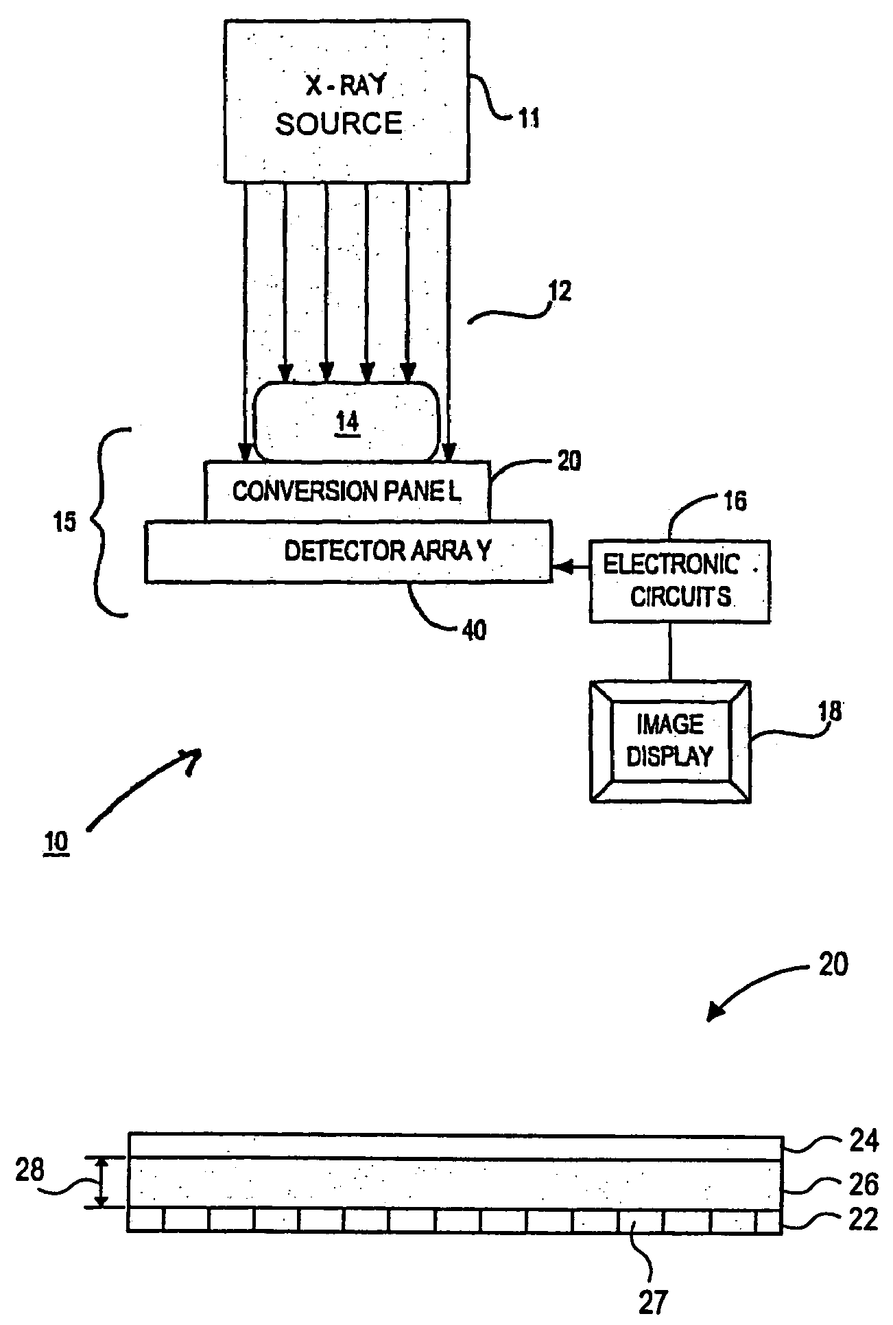
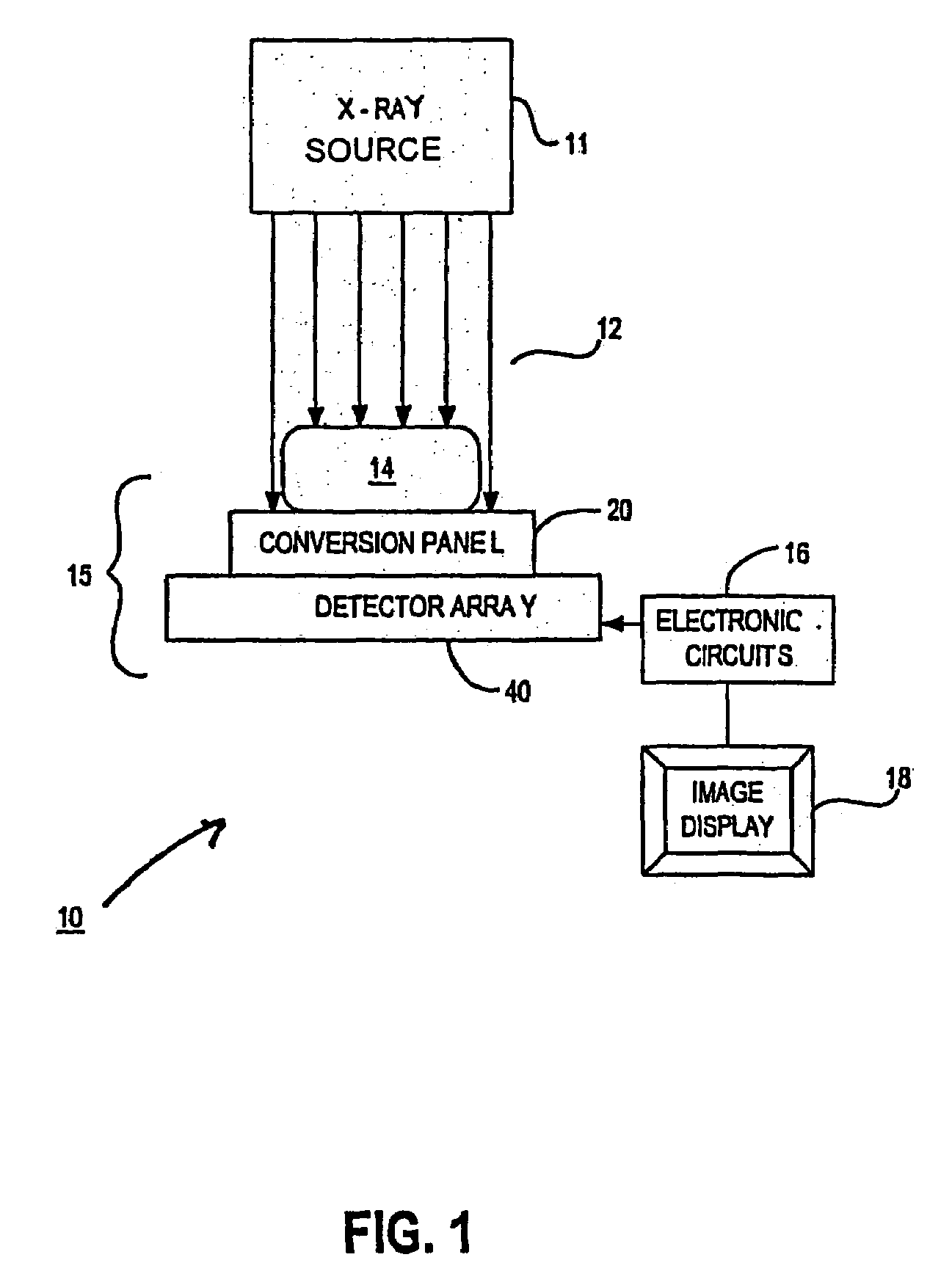
[0017]FIG. 1 is a block diagram schematically illustrating an X-ray imaging system 10 in accordance with an embodiement of the present invention. The X-ray imaging system 10 includes an X-ray source 11 generating X-ray radiation 12 and an X-ray im...


PUM

PropertyMeasurementUnit
thicknessaaaaaaaaaa
thicknessaaaaaaaaaa
thicknessaaaaaaaaaa

Abstract

An X-ray image acquisition apparatus includes a panel having a first electrode, a second electrode, and a photoconductor secured between said first and second electrodes. The photoconductor has a thickness configured to absorb X-ray radiation at a high energy level. The first and second electrodes are configured to create an electric field for transporting charges created in the photoconductor to a pixel unit, thereby allowing charges to be efficiently collected when low or high energy X-ray radiation is used.

Description

FIELD OF THE INVENTION[0001]This invention relates generally to image acquisition and, more specifically, to systems and methods for acquiring an X-ray image.BACKGROUND[0002]X-ray imaging is widely used in various fields of life. For example, X-ray imaging has been a standard medical diagnostic tool for decades.[0003]A typical X-ray image acquisition apparatus suitable for low energy X-rays includes a phosphor X-ray conversion screen and a photo detector array aligned with each other. The phosphor conversion screen generates optical light photons in response to the X-ray radiation. The optical light photons are transmitted to the photo detector array under the conversion screen. The photo detector array generates electric signals in response to the optical light photons. Electronics circuitry coupled to the photo detector array processes the electric signals and generates images.[0004]A typical high energy X-ray image acquisition apparatus includes a copper screen and a Gadolinium O...

Claims


Application Information

Patent Timeline
Patent Type & Authority Patents(United States)
IPC IPC(8): G01N23/00A61B6/00H05G1/60
CPCA61B6/00A61B6/482A61B6/4216
Inventor ZENTAI, GEORGEPARTAIN, LARRY
Owner VARIAN MEDICAL SYSTEMS
Why Eureka
Try Eureka