Device and method for through the scope endoscopic hemostatic clipping

a technology of endoscopic and endoscopic hemostatics, applied in the field of compression clips, can solve the problems of cumbersome operation, time-consuming, and degraded functionality of the device, and achieve the effects of less clips, faster operation, and less bleeding

Inactive Publication Date: 2006-08-22
BOSTON SCI SCIMED INC
View PDF34 Cites 83 Cited by
  • Summary
  • Abstract
  • Description
  • Claims
  • Application Information

AI Technical Summary

Benefits of technology

[0012]The present invention provides medical devices for causing the hemostasis of blood vessels located along the gastrointestinal tract. The goal of the invention is to give the endoscopist a technique and device which: 1) has a success rate in line with the surgical option; 2) is easier to set-up than the Olympus EndoClip; and 3) is easier to deploy than the Olympus EndoClip. The design intent is to eliminate surgery and its associated mortality and morbidity.
[0014]There are several key advantages of the invention disclosed here over existing devices. The device's ability to repeatedly open and close the clip until the desired tissue pinching is accomplished will lead to a quicker procedure, requiring less clips to be deployed, with a higher success rate. In particular embodiments, this higher success rate will be improved even more due to the device's ability to be easily rotated so that the clip legs can be adjusted relative to the bleeding vessel. In particular embodiments, the time required to perform the overall procedure will also be further reduced due to the fact that the device supplied to the endoscopist is completely set up, with the clip already attached to the delivery device, unlike the competitive device. A more robust delivery device may allow a larger, stronger clip to be delivered. Combinations of these features will provide for a device that is easier to use.

Problems solved by technology

This causes the device's functionality to degrade over the course of its use, until it is no longer able to deploy a clip.
The competitive delivery device must be loaded manually, which is cumbersome to the operator and time-consuming, especially in the context of an unplanned emergency procedure.

Method used


Image

  • Device and method for through the scope endoscopic hemostatic clipping
  • Device and method for through the scope endoscopic hemostatic clipping
  • Device and method for through the scope endoscopic hemostatic clipping

Examples


first embodiment

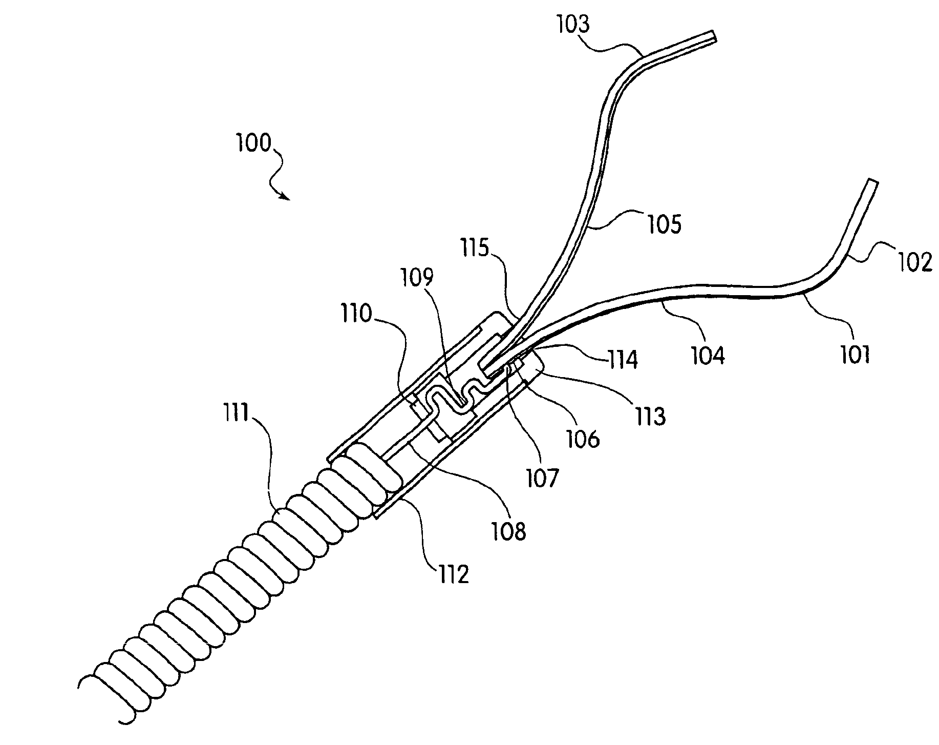
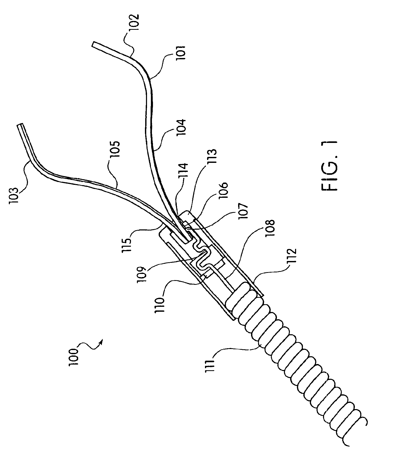
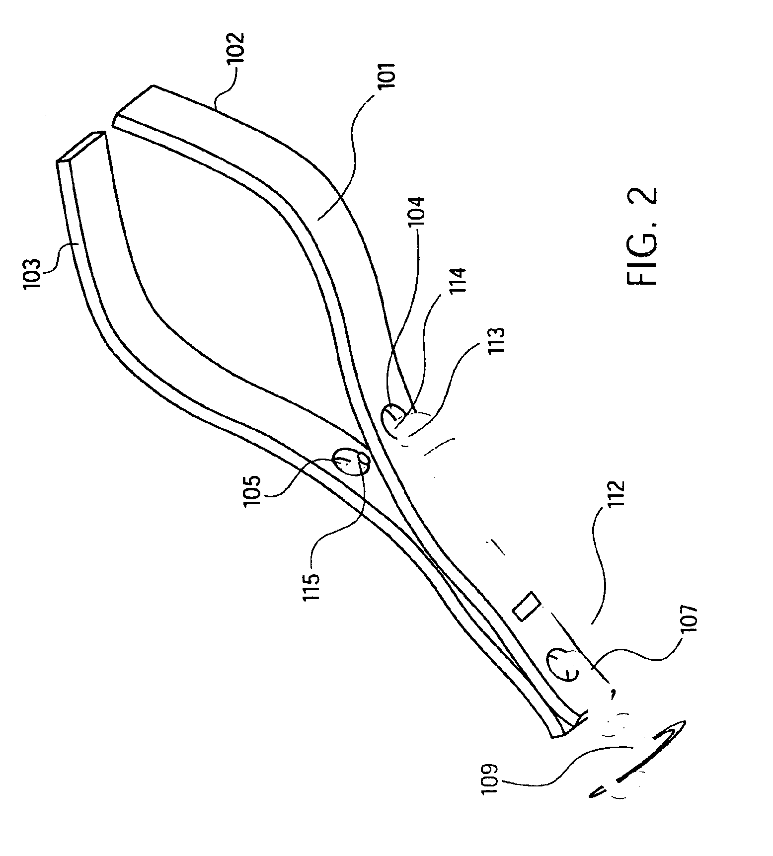
[0067]In the invention as shown in FIG. 1, medical device 100 includes a clip 101 having first clip leg 102 and second clip leg 103. Clip leg 102 has at least one lock hole 104 therein of any suitable shape (e.g. circular, rectangular, square, etc.). Likewise, clip leg 103 has at least one lock hole 105 therein of any suitable shape. Clip 101 is further characterized by a cut-out 106 on the proximal end. J-hook 107 is inserted into cut-out 106. J-hook 107 is formed on the distal terminal end of control wire 108. A retainer release 109 is formed by bends in the control wire 108, the bends formed proximally from the j-hook 107. The control wire 108 is enclosed within sheath 111 proximally from the retainer release 109. Retainer 110 is coupled to control wire 108 and engages lock sleeve 113. Retainer release 109 acts to disengage retainer 110 from lock sleeve 113 when a tensile force applied to control wire 108 is sufficient to cause such disengagement. An outer sleeve 112 is connected...

third embodiment

[0094]FIGS. 18A, 18B, 18C, 18D, 18E, and 18F show an embodiment of a clip which incorporates the natural compressive forces present in a simple elastic band (or o-ring) 1802 to hold the clip legs 1801 in the closed position. FIG. 18A shows two clip legs 1801 in a disassembled state. FIG. 18B shows a clip with the control wire 1803 engaging a second elastic band 1804 to open clip legs 1801. In this embodiment, the control wire 1803 is attached to the proximal end of the clip legs 1801 via a frangible link. In this embodiment, the frangible link is a second elastic band (or o-ring) 1804 that will deform as the control wire 1803 is pulled back. In this embodiment, the clip is housed in the end of a sheath 1806 such that, as the control wire 1803 is pulled back, the second elastic band 1804 delivers an increasing compressive force to the clip legs 1801 proximal to a pin joint 1805, thereby causing the clip legs 1801 distal from the pin joint to open against the compressive force of elas...


PUM


Abstract

Medical device used to cause hemostasis of blood vessels using a clip arrangement delivered to a target region through an endoscope. Method for using the device to cause hemostasis of a blood vessel through an endoscope. Medical device including a reversibly closeable clip, a locking arrangement, a control wire, a sheath, and a handle with an actuating trigger. Through the endoscope, hemostatic clipping device that is fully reversible and lockable. Hemostatic clip that reversibly targets and clips bleeding ulcers.

Description

FIELD OF THE INVENTION[0001]The present invention relates to compression clips, and more specifically, to compression clips used to cause hemostasis of blood vessels located along the gastrointestinal tract delivered to a target site through an endoscope.BACKGROUND[0002]Gastrointestinal (“GI”) bleeding is often associated with peptic ulcer disease (PUD) and can be fatal if not treated immediately. Hemorrhaging is the most dangerous procedure with which a Gastro-Intestinal Endoscopist has to deal. It is his / her only unplanned, emergency procedure where time is critical in determining the outcome. It is also the one problem the Endoscopist faces that is generally not an outpatient procedure. A bleeding PUD can be a critical clinical event as there is internal hemorrhaging. Ulcers are classified from clean to active spurting bleeding. The most worrisome are active bleeders and visible vessels. Untreated visible vessels are likely to bleed.[0003]Suspected bleeding PUD patients can be di...

Claims


Application Information

Patent Timeline
Patent Type & Authority Patents(United States)
IPC IPC(8): A61B17/10A61B1/00A61B17/12A61B17/00A61B17/122A61B17/128A61B19/00
CPCA61B17/122A61B17/1285A61B17/083A61B2017/12004A61B2019/307A61B2017/00477A61B2090/037A61B2090/3916A61B2017/0034A61B90/03A61B17/1227A61B2017/00778A61B2017/00818
Inventor ADAMS, MARK L.DURGIN, RUSSELL F.TURTURRO, VINCENTGRANT, JUSTINMAY, NORMANSULLIVAN, III, ROY H.
Owner BOSTON SCI SCIMED INC
Why Eureka
Try Eureka