Methods and compositions for the visualization of cellular organelles using tetrazolium salts

Inactive Publication Date: 2006-09-05
KONG QINGZHONG
View PDF12 Cites 2 Cited by
  • Summary
  • Abstract
  • Description
  • Claims
  • Application Information

AI Technical Summary

Benefits of technology

[0014]The inventor has found that enzymes, such as dehydrogenases, are highly expressed in various tissues, and cell lines, particularly in cancerous cells. Except for mitochondrion, this enzyme is widely expressed in other structures of the cells such as, but not limited to, centrosomes, microtubules, endoplasmic reticulum, flagella, nuclear envelopes, lysosomes and other structures as mentioned above. When crystallizing agents, such as tetrazolium salts, are present, cellular enzymes, mainly succinate dehydrogenase (SDH), which is usually anchored on the cellular structures, will reduce the tetrazolium salts into related crystals that can be visualized under proper conditions. Additionally, the formation of crystals may act as a restraint to the separation of the duplicable organelles and to the movement of cytoskeletons.
[0015]Based on this discovery, the present invention features a method for visualizing cellular organelles and / or cytoskeletons, in tissue or in cell suspension, by treating tissue or cells with one or more crystallizing agents. The method includes obtaining a tissue sample or a fluid sample containing multiple cells from a variety of sources such as, but not limited to mammalian, microorganisms, and cancers; then fixing the tissue sample or cultured cell sample; contacting the tissue sample or cell sample with a crystallizing agent under conditions that permit the formation of visible crystals through the reduction of said crystallizing agent by the enzymes located in said cellular organelles and / or cytoskeletons in said cells or tissues; and then visualizing cellular organelles and / or cytoskeletons having crystals formed on them. The method allows for easy and quick visualization of cellular organelles and / or cytoskeletons that are usually observed through complex immunofluorescence staining or under electronic microscope.

Problems solved by technology

These techniques have been proved to be very costly, poorly reproducible, time consuming, and requiring of very strict conditions.
Particularly, these methods can not be used to demonstrate the dynamic states of cells.

Method used


Image

  • Methods and compositions for the visualization of cellular organelles using tetrazolium salts
  • Methods and compositions for the visualization of cellular organelles using tetrazolium salts
  • Methods and compositions for the visualization of cellular organelles using tetrazolium salts

Examples


example 1

Preparation of Cells from Fresh Tissues for Cellular Organelle Visualzation

[0054]Cells suspensions were removed from surgical resection specimens of intracranially or subcutaneously established gliosarcomas in rats by removing samples (0.5-1.0 cm square) of tissue and then mincing the samples with a razor blade in PBS at room temperature. Minced tissue was then washed in PBS and resuspended in 2 ml aliquot of PBS containing 2.0 U / ml of collagenase and 0.2 U / ml DNase, and then incubated for 2 hours at room temperature. During the incubation, the samples were rotated end over end. Following collagenase and DNase digestion, the samples were strained on a 100 micrometer nylon filter (Nytex, Small Parts, Inc., Miami Lakes, Fla.), pelleted, washed in PBS, by sequential centrifugation at 325 G. Then they were transferred into a flask for culture as described in Examples 3-6 (below). Cells can also be obtained from fresh tissue, peripheral blood, bone marrow, and lymph nodes and treated in ...

example 2

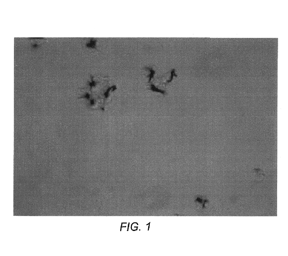
Visualization of Centrosomes of SHP77 (SCLC) Cells following Treatment with TV

[0055]9L gliosarcoma tumor cells (4000) obtained by the method described in example 1 were seeded in 48 well plate one day before being exposed to tetrazolium violet (TV, a substrate of SDH) at the concentration of 50 μg / ml. The Modified DMEM media was used in this example. As shown in FIG. 1, centrosomes, usually one in each cell (J, #1-6), were crystallized at the poles, with the cell viability being significantly reduced (see data in Example 1).

[0056]After being treated under the same condition, the centrosome of CNS1 tumor cells were all crystallized (not shown).

example 3

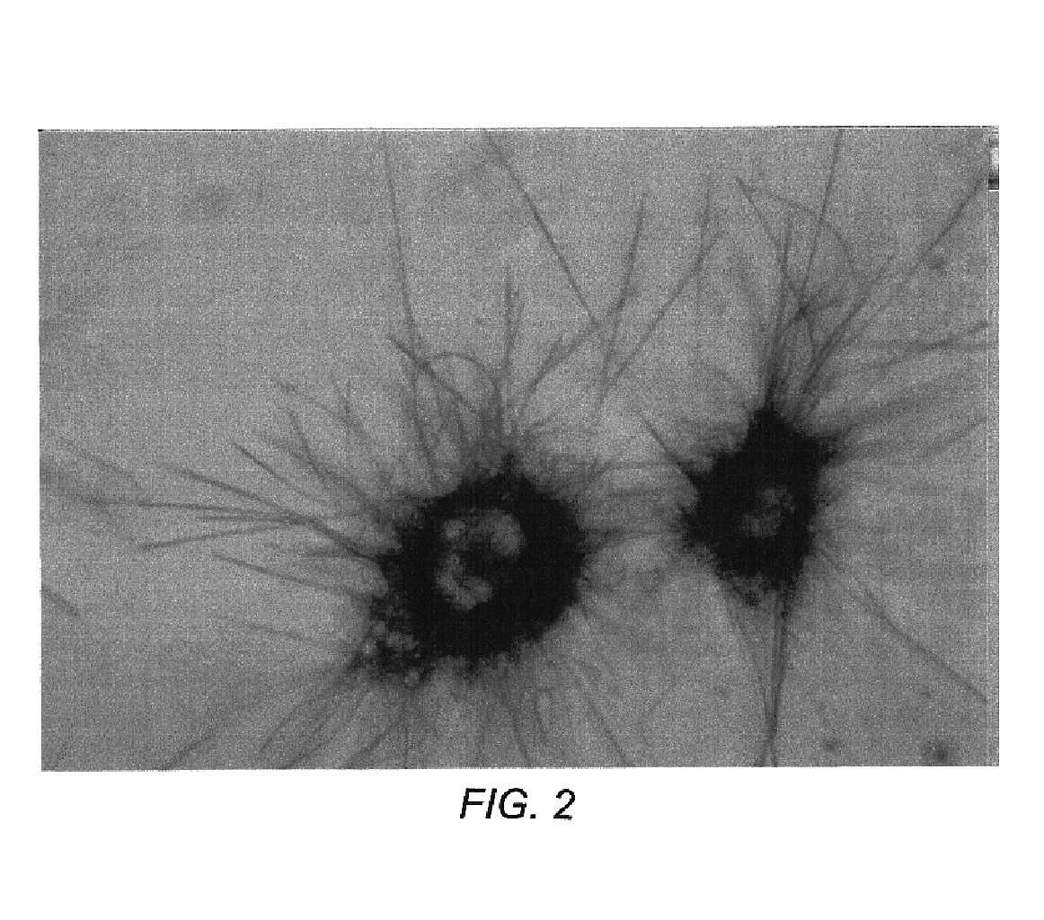
Visualization of Microtubules of 9L Gliosarcoma Cancer Cells following Treatment with MTT

[0057]9L cancer cells were exposed to MTT (50 ug / ml) at the same condition as described in Example 2. The tumor cells (4000) were seeded one day before being treated with MTT. As can be seen in FIG. 2, the microtubules and nuclear envelopes were frozen by the crystals formed on them.


PUM


Abstract

The invention provides a biochemical method for visualizing cellular organelles (such as centrosome) and / or cytoskeletons (such as microtubules) through the use of crystallizing agents (such as tetrazolium salts), a kit containing crystallizing agents adapted for such uses, and to methods particularly for detecting neoplastic cells in a tissue sample, suspension, or a fluid sample by examining the cells for abnormalities of cellular organelles (such as centrosome) and / or cytoskeletons (such as microtubules) in the number and shapes.

Description

CROSS REFERENCE TO RELATED APPLICATIONS[0001]The present application is a divisional of U.S. patent application Ser. No. 09 / 687,342, now U.S. Pat. No. 6,368,818, filed Oct. 12, 2000, entitled “METHODS AND COMPOSITIONS FOR THE VISUALIZATION OF CELLULAR ORGANELLES USING TETRAZOLIUM SALTS,” which is incorporated herein by this reference.FIELD OF THE INVENTION[0002]The invention relates to cell biology and oncology. More specifically, this invention relates to the methods for visualizing cellular organelles (such as a centrosome) and / or cytoskeletons (such as microtubules) through the use of crystallizing agents (such as tetrazolium salts), to a kit containing crystallizing agents adapted for such use, and to methods particularly for detecting neoplastic cells in a tissue sample, suspension, or a fluid sample by examining the cells for abnormalities in the size, number and shape of cellular organelles (such as a centrosome) and / or cytoskeletons (such as microtubules).BACKGROUND OF THE I...

Claims


Application Information

Patent Timeline
IPC IPC(8): C07D257/04C07D257/10C07D403/00C07D487/00A61K31/41
CPCA61K31/41
Inventor KONG
Owner KONG QINGZHONG
Why Eureka
Try Eureka