

FXIII detection for verifying serum sample and sample size and for detecting dilution
a technology of fxiii and detection method, which is applied in the field of quality control of clinical laboratory test procedures and instruments, can solve the problem that the disease state has a sufficiently small effect on the concentration of this subunit, and achieve the effect of rare deficiency
- Summary
- Abstract
- Description
- Claims
- Application Information
AI Technical Summary
Benefits of technology
Problems solved by technology
Method used
Image

Examples
example 1
[0038]This example provides the results of sequential immunoassays performed on microparticles to illustrate the ability of the present invention to distinguish plasma from serum by analyzing for the presence of dissociated subunit a of human Factor XIII protein.
[0039]The assays were sandwich-type immunoassays and the microparticles were 7.1 μm magnetic microparticles. A portion of the microparticles was coated with polyclonal anti-FXIIIa2b2 antibody and a second portion was coated with polyclonal anti-FXIIIa antibody (as capture antibodies). The polyclonal anti-FXIIIa2b2 antibody was specifically reactive toward the tetramer, while the polyclonal anti-FXIIIa antibody was reactive toward both free and bound a subunit and non-reactive toward free b subunit. A different polyclonal anti-FXIIIa2b2 antibody was used as the label antibody for the tests on both portions. Assays on the first portion of microparticles thus indicated the presence of the tetramer and any dissociated subunits, ...
example 2
[0047]This example provides the results of ELISAs (enzyme-linked immunosorbent assays) to illustrate the ability of the present invention to distinguish plasma from serum, this time using monoclonal antibody specific for the tetramer as the capture antibody in all cases, and polyclonal antibody specific for subunit a and polyclonal antibody specific for subunit b in separate assays as the detection antibody.
[0048]The assays were performed on coated microplates, and as in Example 1, two assays were performed on serum and two on plasma. To coat the plates, the capture antibody was diluted 1 / 100 with 50 mM carbonate buffer, pH 9.6, and added to the microplate wells in volumes of 100 μL per well, followed by incubation overnight at 4° C. per well. The wells were emptied immediately before use and blocking buffer was added, followed by four washes with phosphate-buffered saline (PBS) and Tween surfactant. Plasma and serum samples were diluted 1 / 400 with PBS and added to the wells. Detect...
example 3
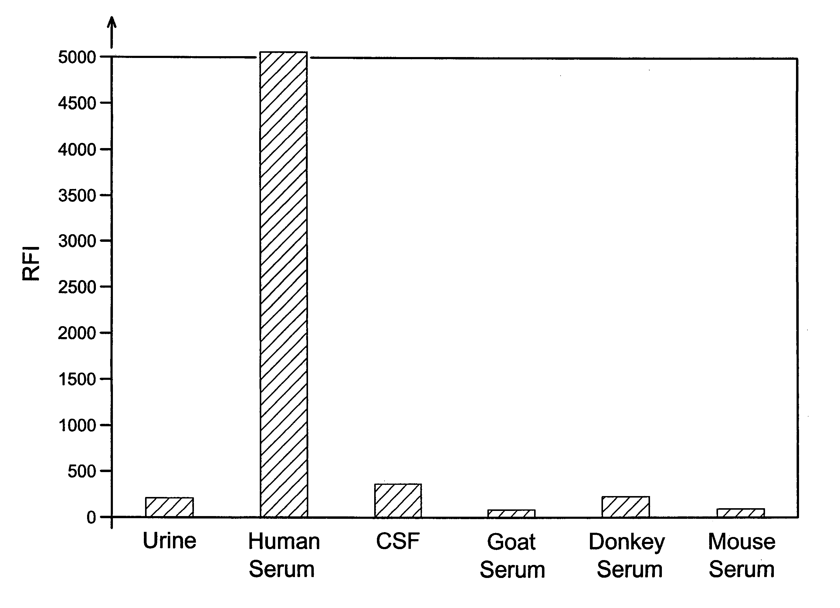
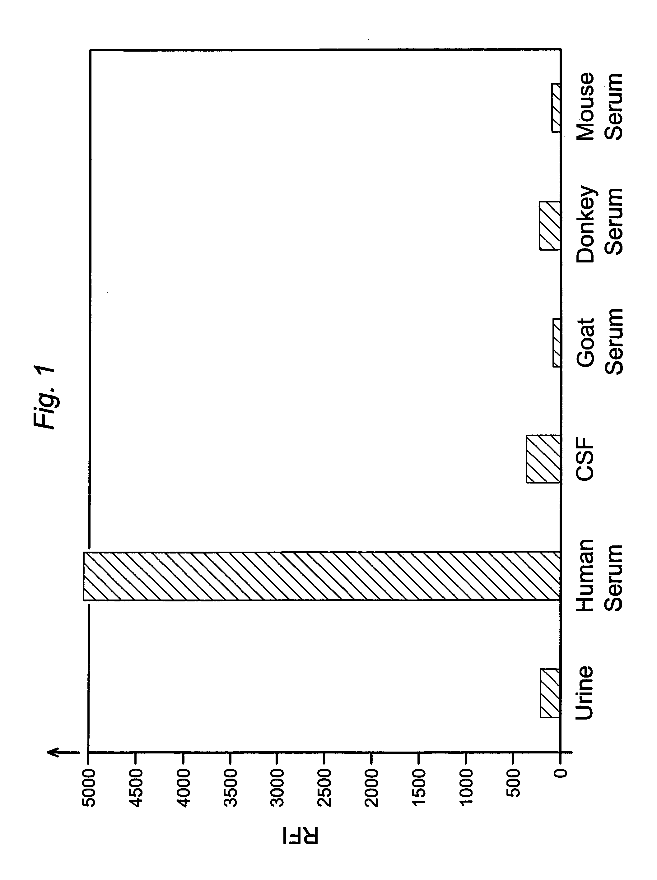
[0052]This example compares the results of assays for the FXIII tetramer in human urine, human cerebrospinal fluid, and serum from various species.
[0053]The assays were sandwich-type immunoassays performed on magnetic microparticles as in Example 1, using monoclonal anti-human FXIIIa2b2 antibody as the capture antibody and polyclonal anti-human FXIIIa2b2 antibody conjugated with phycoerythrin as the label antibody. The particles, which were 8.0 μm in diameter, were coated with capture antibody in the manner described in Example 1, and antibody-label conjugates were likewise prepared as described in Example 1. The particles were suspended in wash buffer to a concentration of 47 μg / mL, and 100 μL aliquots of the particle suspensions were mixed with 100 μL each of human serum, cerebrospinal fluid, urine, donkey serum, mouse serum, and goat serum. The resulting particle / sample suspensions were incubated on a shaker (1,100 rpm) at room temperature for fifteen minutes. Unbound and contami...
PUM
| Property | Measurement | Unit |
|---|---|---|
| concentration | aaaaa | aaaaa |
| concentration | aaaaa | aaaaa |
| weight percent | aaaaa | aaaaa |
Abstract
Description
Claims
Application Information