Elastographic imaging of in vivo soft tissue

Active Publication Date: 2007-01-23
WISCONSIN ALUMNI RES FOUND
View PDF17 Cites 98 Cited by
  • Summary
  • Abstract
  • Description
  • Claims
  • Application Information

AI Technical Summary

Benefits of technology

[0008]The present inventors have recognized that compression of the tissue may be done by a probe inserted percutaneously into the tissue

Problems solved by technology

While elastography has great promise, in vivo elastography of soft tissue structures such as the liver and other abdominal

Method used


Image

  • Elastographic imaging of in vivo soft tissue
  • Elastographic imaging of in vivo soft tissue
  • Elastographic imaging of in vivo soft tissue

Examples


Embodiment Construction

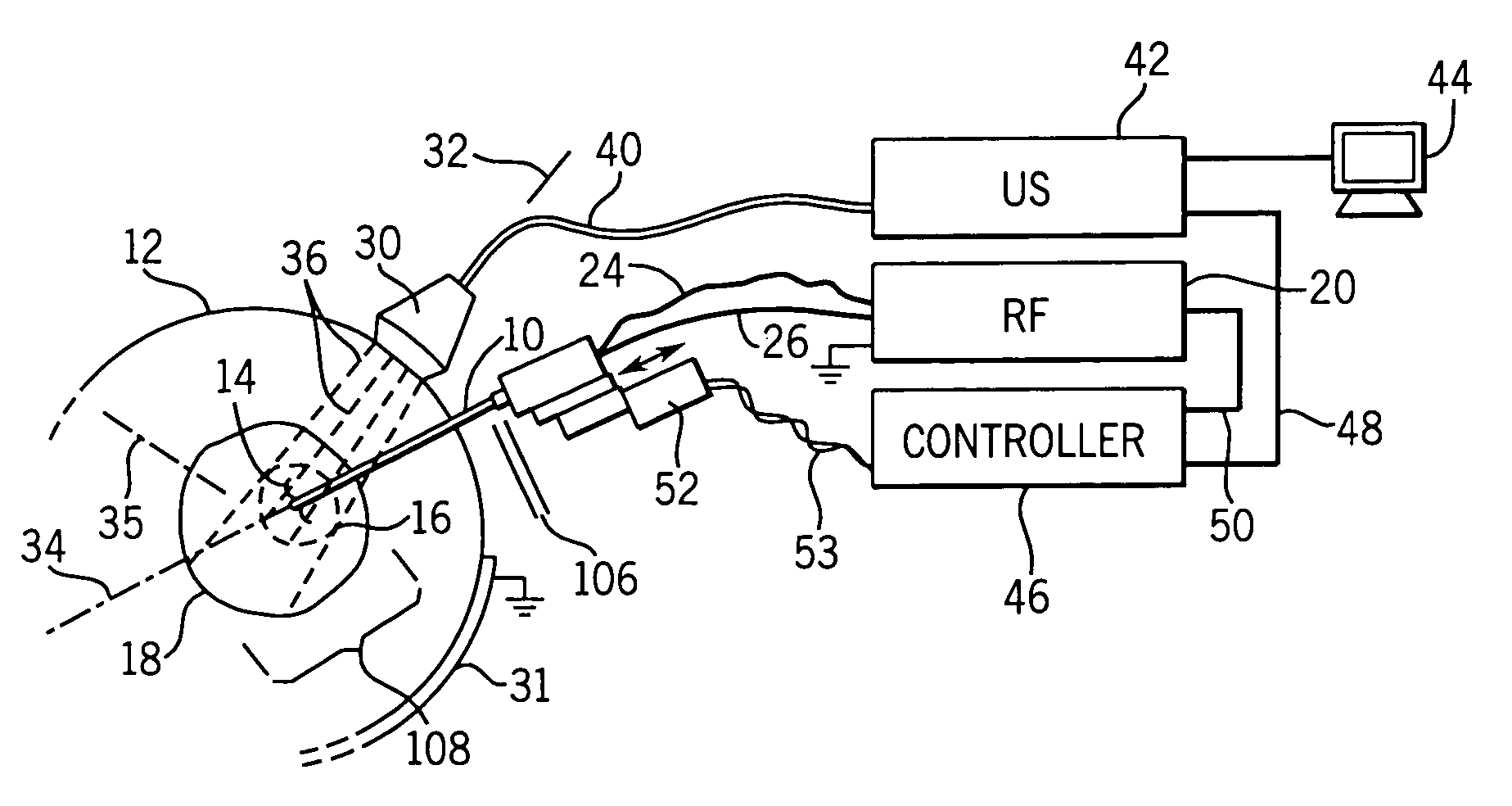
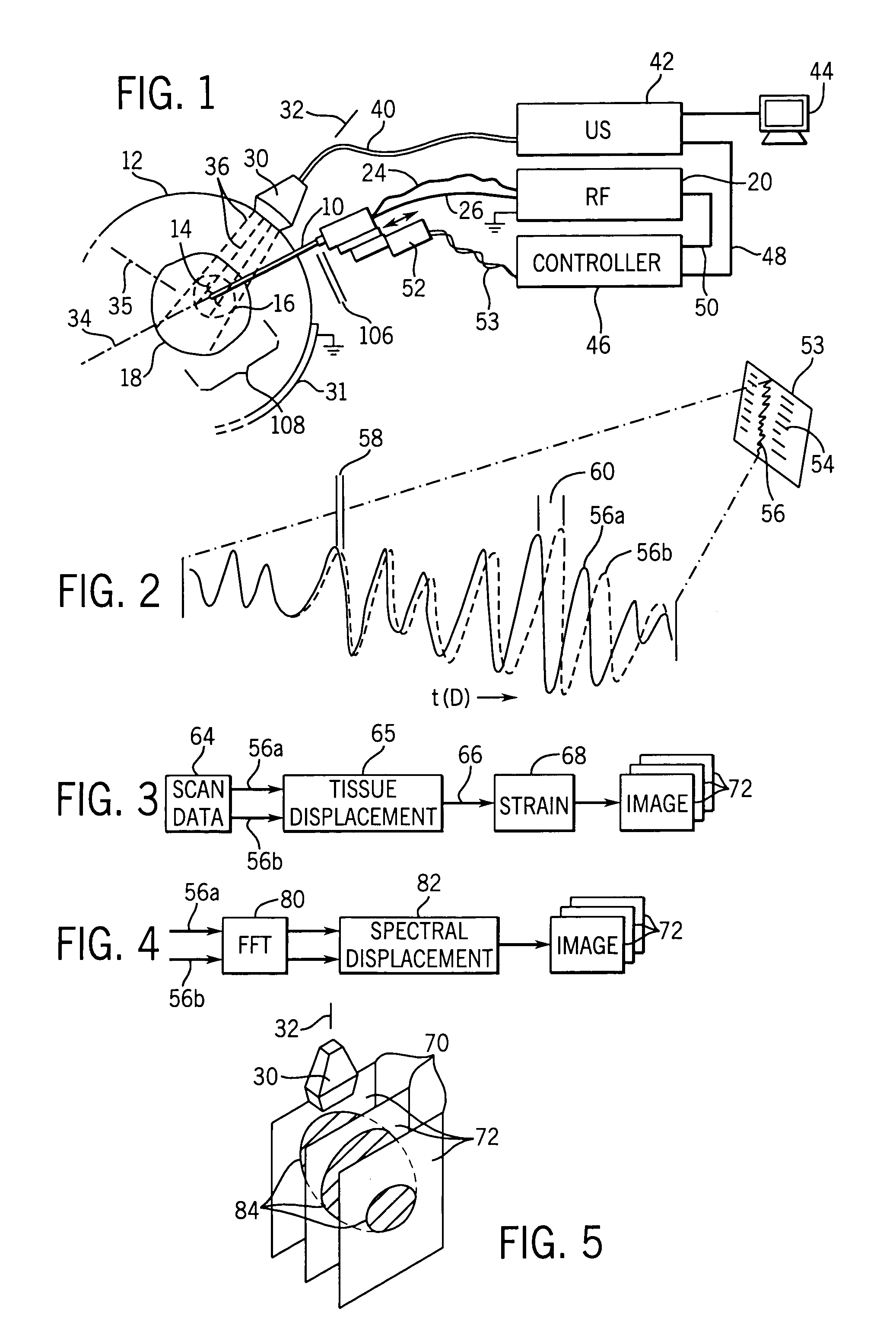
[0042]Referring now to FIG. 1, an RF ablation probe 10 may be inserted percutaneously into a patient 12 to have its tip located at an ablation region 16 within an organ 18 such as the liver.

[0043]Extensible electrode tines 14, at the tip of the probe 10, may grip the tissue of the ablation region and provide a greater area of electrical contact to conduct ablative current from an RF source 20. Electrical energy from the RF source 20 is conducted through an insulated shaft of the probe 10 to the conductive tines 14 where ionic heating of the tissue kills tumor tissue. A large-area grounding pad 31 placed on the patient's skin provides a return path for this current. The tines 14 may include thermocouples for temperature measurements.

[0044]RF ablation probes 10 of this kind having extensible tines and thermocouple sensors are well known in the art and readily available. The RF 20 source may be a Rita Model 30 electrosurgical device manufactured by Rita Medical Systems Inc., Mountain V...


PUM


Abstract

Elastographic images provide visualization in two or three dimensions of RF ablation lesions to guide in the ablation process. Compression may be applied using the RF probe. A similar technique may be applied to in vivo imaging of soft tissue without ablation.

Description

CROSS-REFERENCE TO RELATED APPLICATIONS[0001]--STATEMENT REGARDING FEDERALLY SPONSORED RESEARCH OR DEVELOPMENT[0002]--BACKGROUND OF THE INVENTION[0003]The present invention relates to medical imaging, and in particular to methods of in vivo elastographic imaging.[0004]Elastography is a new imaging modality that reveals the stiffness properties of tissues, for example, axial strain, lateral strain, Poisson's Ratio, Young's Modulus or other common stiffness measurements. The measurements provide a two-dimensional array of data in which array location corresponding to tissue locations in an image plane. The array of data may be mapped to a gray scale to form a picture.[0005]In “quasi-static” elastography, two images of the tissue are obtained. The first image forms a base line of the tissue in an unstressed or relaxed state or under a small pre-compression. The second image is obtained with the tissue under compression. Displacement of the tissue between the two images is used to deduc...

Claims


Application Information

Patent Timeline
IPC IPC(8): A61B8/00A61B8/08A61B8/14G01S7/52
CPCA61B5/0048A61B5/015A61B8/0833A61B8/14A61B8/485A61B8/483A61B18/14A61B2018/00577G01S7/52046A61B8/466A61B8/5223
Inventor VARGHESE, TOMYZAGZEBSKI, JAMES A.TECHAVIPOO, UDOMCHAICHEN, QUAN
Owner WISCONSIN ALUMNI RES FOUND
Why Eureka
Try Eureka