
Cellular surgery utilizing confocal microscopy

a confocal microscopy and cellular technology, applied in the field of cellular surgery utilizing confocal microscopy, can solve the problems of not using confocal microscopy for tissue imaging, not being able to treat cells in confocal systems, and not being able to provide treatment of cells of in-vivo tissue of patients

Inactive Publication Date: 2007-03-13
CALIBER IMAGING & DIAGNOSTICS
View PDF27 Cites 9 Cited by
  • Summary
  • Abstract
  • Description
  • Claims
  • Application Information

AI Technical Summary

Benefits of technology

[0006]Accordingly, the principal object of the present invention is to provide an improved system for generating confocal images of in-vivo tissue which enables surgical treatment of tissue being imaged.
[0007]A further object of the present invention is to provide an improved system for generating confocal images of in-vivo tissue which enables surgical treatment either to be localized to a small region of tissue being imaged, or to be non-localized over a region of tissue including that small region of tissue.
[0008]Another object of the present invention is to provide an improved system for generating through confocal optics images of in-vivo tissue which enables laser surgical treatment of the tissue being imaged, allows for evaluating the effectiveness of such treatment by simultaneously or sequentially imaging the treated tissue, and for modifying the operating parameters of the laser and / or confocal optics in subsequent treatments of the tissue.
[0009]Briefly described, the present invention embodies a system including a laser for producing a laser beam, and confocal optics for scanning and focusing the beam in tissue and collecting returned light from the tissue. A detector is provided which confocally detects the returned light and produces signals in accordance with the detected returned light representing confocal images. Responsive to the signals, such confocal images are visualized on a display. The system further includes a programmed controller for enabling the operator to select one or more cells of the tissue in the visualized confocal images for surgical treatment. The controller operates the laser and confocal optics in a first mode at a first set of operating parameters to treat the tissue when the confocal optics focus the laser beam at least one region associated with the selected cells in the tissue, but at all other times operates the laser and confocal optics in a second mode at a second set of operating parameters which does not damage the tissue.

Problems solved by technology

These confocal systems although useful for examination of lesions or other diseased tissue have no capability for treatment of cells, such as, for example, to cause thermolysis, photolysis, or ablation of imaged cells.
This device however does not use confocal microscopy for tissue imaging and does not provide treatment of cells of in-vivo tissue of a patient.

Method used


Image

  • Cellular surgery utilizing confocal microscopy
  • Cellular surgery utilizing confocal microscopy
  • Cellular surgery utilizing confocal microscopy

Examples


Embodiment Construction

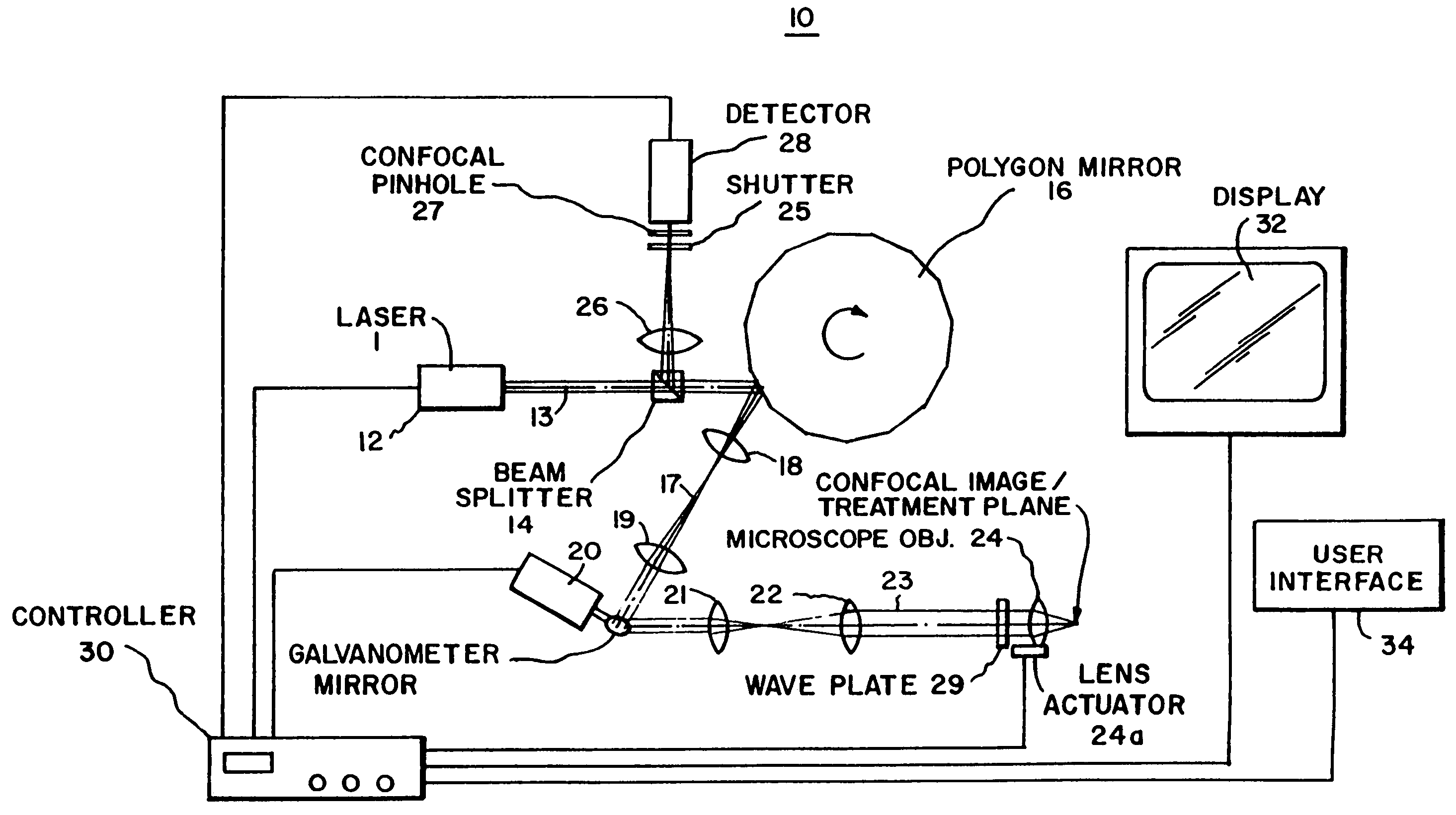
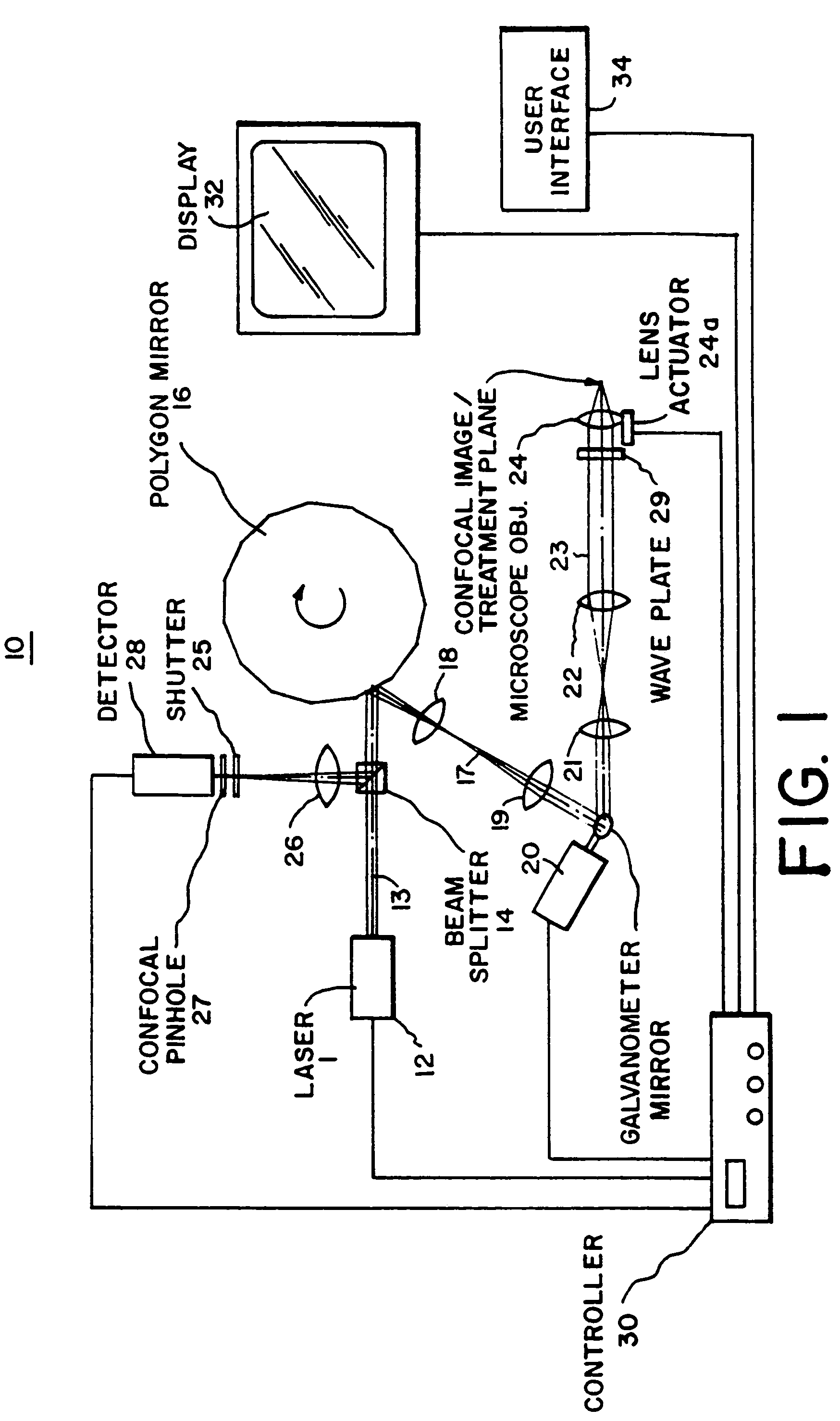
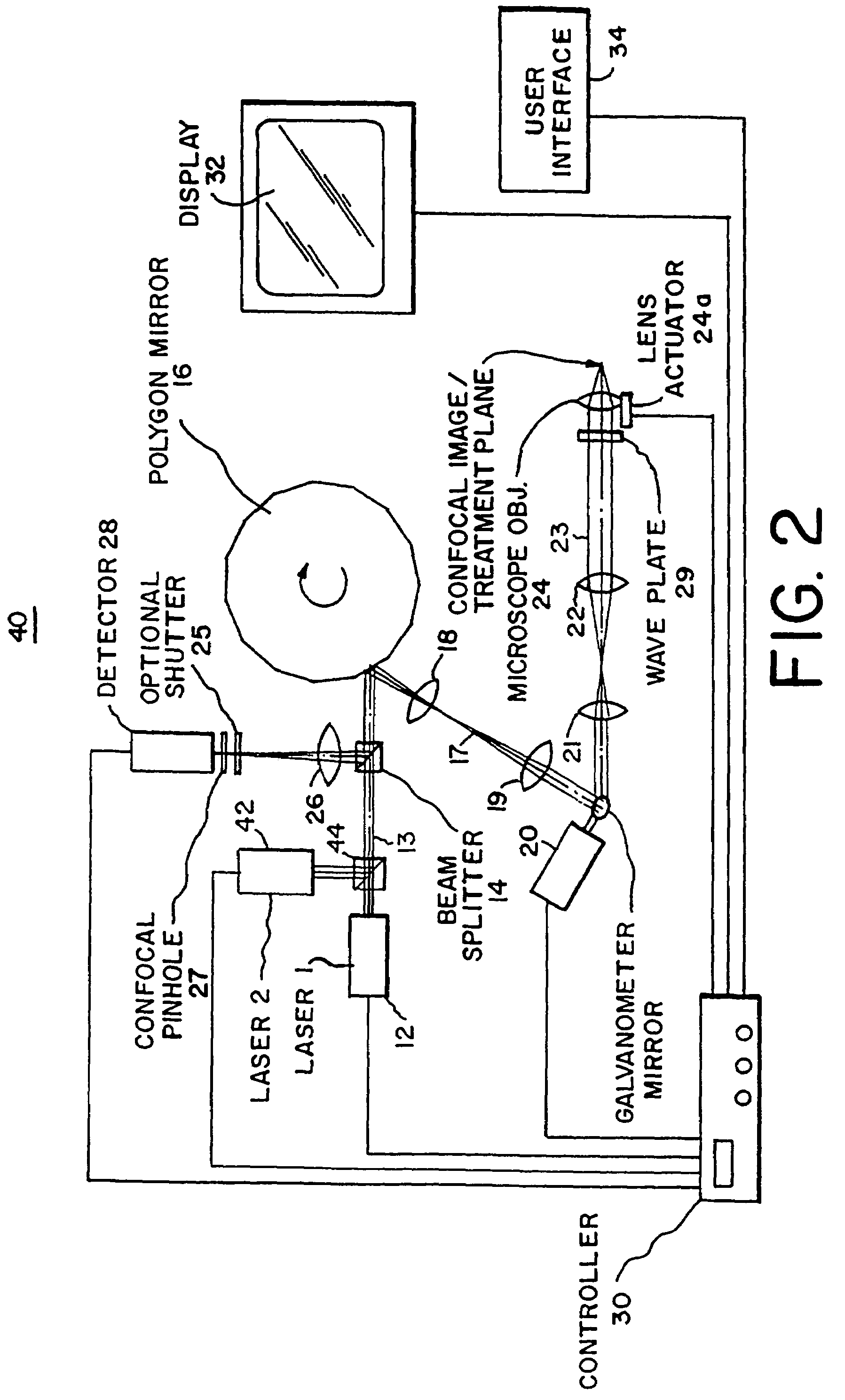
[0017]Referring now to FIG. 1, a system 10 of the present invention is shown. System 10 includes a first laser 12 (Laser 1) for producing light (a laser beam) at an infrared wavelength along a path 13 through beam-splitter 14 onto a rotating polygon mirror 16. Polygon mirror 16 has a plurality of mirror facets to reflect the beam from laser 12 at varying angles responsive to the rotation of mirror 16, i.e., to repeatedly scan the beam. The reflected beam from rotating polygon mirror 16 travels along a path 17 through relay and focusing lenses 18 and 19 onto a galvanometer mirror 20. Lenses 18 and 19 image the beam reflected by the polygon mirror facet onto galvanometer mirror 20. Galvanometer mirror 20 reflects the beam incident thereto at a controlled angle through lenses 21 and 22 along a path 23 to an objective focusing lens 24. Lenses 21 and 22 image the beam reflected by galvanometer mirror 20 onto objective lens 24. A quarter-wave plate 29 is provided in path 23 between lens 2...


PUM


Abstract

An improved system for cellular surgery which includes a laser for producing a laser beam, and confocal optics for scanning and focusing the laser beam in tissue and generating confocal images of the tissue in accordance with returned light from the tissue. The confocal images are visualized on a display. The system includes a controller for enabling the operator to select one or more cells of the tissue in the displayed confocal images for surgical treatment. The controller operates the laser and confocal optics in a first mode to treat the tissue when the confocal optics focus the laser beam at least one region associated with the selected cells in the tissue, but at all other times operates the laser and confocal optics in a second mode which does not damage the tissue. The treatment may be localized to concentrate the energy of the laser to the region including the selected cell or cells, or the treatment may be non-localized to distribute the energy of the laser to the region which includes the selected cell(s) and also the cells of the tissue surrounding such selected cell(s). In another embodiment, an apparatus is provided having a confocal imaging system, which focuses a first laser beam through confocal optics to tissue and provides confocal images of the tissue, and a treatment system which focuses a second laser beam through the confocal optics coaxial with the first laser beam for treating at one or more selected locations in the imaged tissue.

Description

DESCRIPTION [0001]This application is a divisional of application Ser. No. 09 / 438,065, filed Nov. 10, 1999, now U.S. Pat. No. 6,668,186, which is a continuation of application Ser. No. 09 / 044,355, filed Mar. 19, 1998, now U.S. Pat. No. 5,995,867, which claims the benefit of priority from U.S. Provisional Application Ser. No. 60 / 041,050, filed Mar. 19, 1997.FIELD OF THE INVENTION[0002]The present invention relates to a system (method and apparatus) for cellular surgery utilizing confocal microscopy, and relates particularly to, a system for cellular surgery which provides for confocal imaging of tissue and treatment of one or more cells of the tissue being imaged. Cellular surgery is herein defined as surface or subsurface excision, ablation, thermolysis, photo-drug activation, or photo-chemical or photo-acoustical changes, on a region of tissue characterized by one or more individual cells.BACKGROUND OF THE INVENTION[0003]Confocal microscopy involves scanning tissue to produce micro...

Claims


Application Information

Patent Timeline
IPC IPC(8): A61B6/00G02B21/36A61B5/00A61B18/20A61B19/00A61N5/06G02B21/00G02B26/12
CPCA61B18/20G02B26/12G02B21/0012G02B21/0028A61B18/203A61B19/5223A61B2018/00452A61B2562/0242A61B2018/00636A61B90/20
Inventor ZAVISLAN, JAMES M.GREENWALD, ROGER J.
Owner CALIBER IMAGING & DIAGNOSTICS
Learn More