Antibodies to JTT-1 protein and cells secreting such antibodies

a technology of jtt-1 and antibodies, applied in the field of mammals' new cell surface molecules, can solve problems such as clonal anergy or unresponsiveness

Inactive Publication Date: 2007-03-27
JAPAN TOBACCO INC
View PDF39 Cites 45 Cited by
  • Summary
  • Abstract
  • Description
  • Claims
  • Application Information

AI Technical Summary

Benefits of technology

[0046]It was also found that the monoclonal antibody against “human JTT-1 antigen” significantly proliferated human peripheral blood lymphocytes, and that the proliferation was further enhanced in the co-presence of a monoclonal antibody against CD3 constituting a TcR / CD3 complex on T cells, which receives the primary signal essential for T cell activation from antigen presenting cells, indicating that “JTT-1 antigen” was a cell surface molecule involved in signal transmission into lymphocytes.
[0178]The fusion polypeptide of the present invention has the advantage that the fusion polypeptide can be purified extremely easily by using affinity column chromatography using the property of protein A, which binds specifically to the immunoglobulin fragment because the fusion polypeptide of the present invention has a portion of a constant region (for example Fc) of an immunoglobulin such as IgG as mentioned above as a fusion partner. Moreover, since various antibodies against the Fc of various immunoglobulin are available, an immunoassay for the fusion polypeptides can be easily performed with antibodies against the Fc.
[0207]Examples of a method for introducing a plasmid into a host are calcium chloride method, calcium chloride / rubidium chloride method described in Molecular Cloning, A Laboratory Manual (second edition, Cold Spring Harbor Laboratory, p. 1.74 (1989)), and electroporation method. Phage vectors can be introduced into host cells by, for example, a method in which the phage DNAs are introduced into grown hosts after in vitro packaging. In vitro packaging can be easily performed with a commercially available in vitro packaging kit (for example, a product from Stratagene or Amersham).
[0217]The recombinant vector can easily be prepared by ligating the DNA encoding the polypeptide of the present invention with a vector for recombination available in the art (plasmid DNA and bacteriophage DNA) by the usual method. Specific examples of the vectors for recombination used are E. coli-derived plasmids such as pBR322, pBR325, pUC12, pUC13, and pUC19, yeast-derived plasmids such as pSH19 and pSH15, and Bacillus subtilis-derived plasmids such as pUB110, pTP5, and pC194. Examples of phages are a bacteriophage such as λ phage, and an animal or insect virus (pVL1393, Invitrogen) such as a retrovirus, vaccinia virus, and nuclear polyhidrosis virus.

Problems solved by technology

However, the TcR / CD3 complex-mediated primary signal alone cannot activate T cells sufficiently and leads to unresponsiveness or clonal anergy, so that the cells can not react with any stimulation received thereafter.

Method used


Image

  • Antibodies to JTT-1 protein and cells secreting such antibodies
  • Antibodies to JTT-1 protein and cells secreting such antibodies
  • Antibodies to JTT-1 protein and cells secreting such antibodies

Examples


example 1

Preparation of Monoclonal Antibodies

[0336]Antibody-producing hybridomas were prepared according to the method of Köhler et al. (Omori et al., Blood, 81:101–111, 1993), and monoclonal antibodies were prepared according to the method of Kannagi et al. (Handbook of Experimental Immunology, 4:117.21–117.21, 1986).

[0337]First, rat thymoma cell line FTL435 cells were administered as an immunizing antigen to BALB / c mice into their footpad in an amount of 107 cells / mouse at intervals of 0, 7, 14, and 28 days. The mixture of the antigen with Freund's complete adjuvant was administered only in the first immunization. Two days after the last immunization, the lymph nodes of the mice were taken out and fused with mouse myeloma cells PAI (JCR No. B0113; Stocker et al., Res. Disclosure, 217:155, 1982) by the usual method to obtain many hybridomas producing monoclonal antibodies.

example 2

Screening of Hybridomas and Characterization of Monoclonal Antibodies

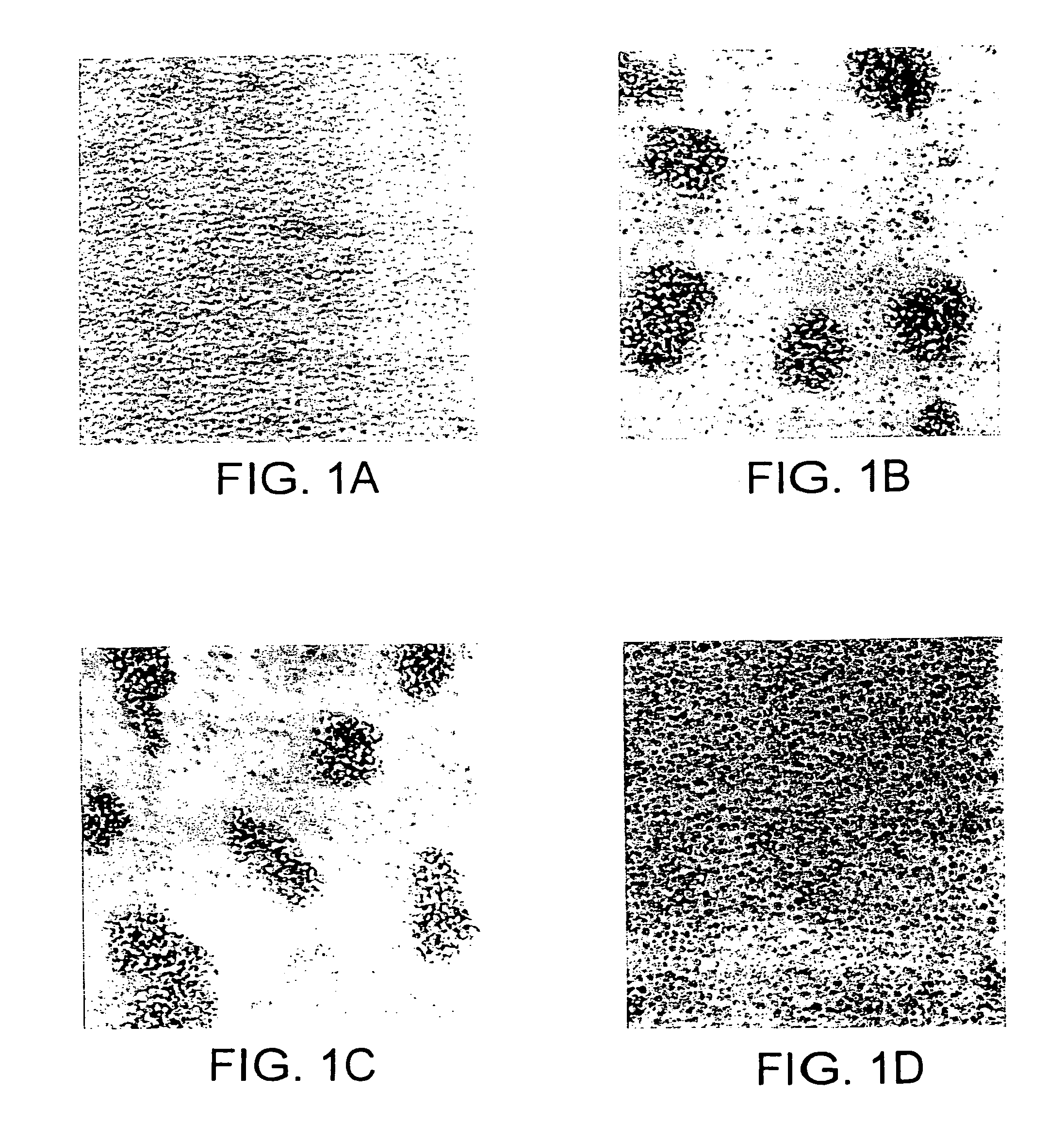
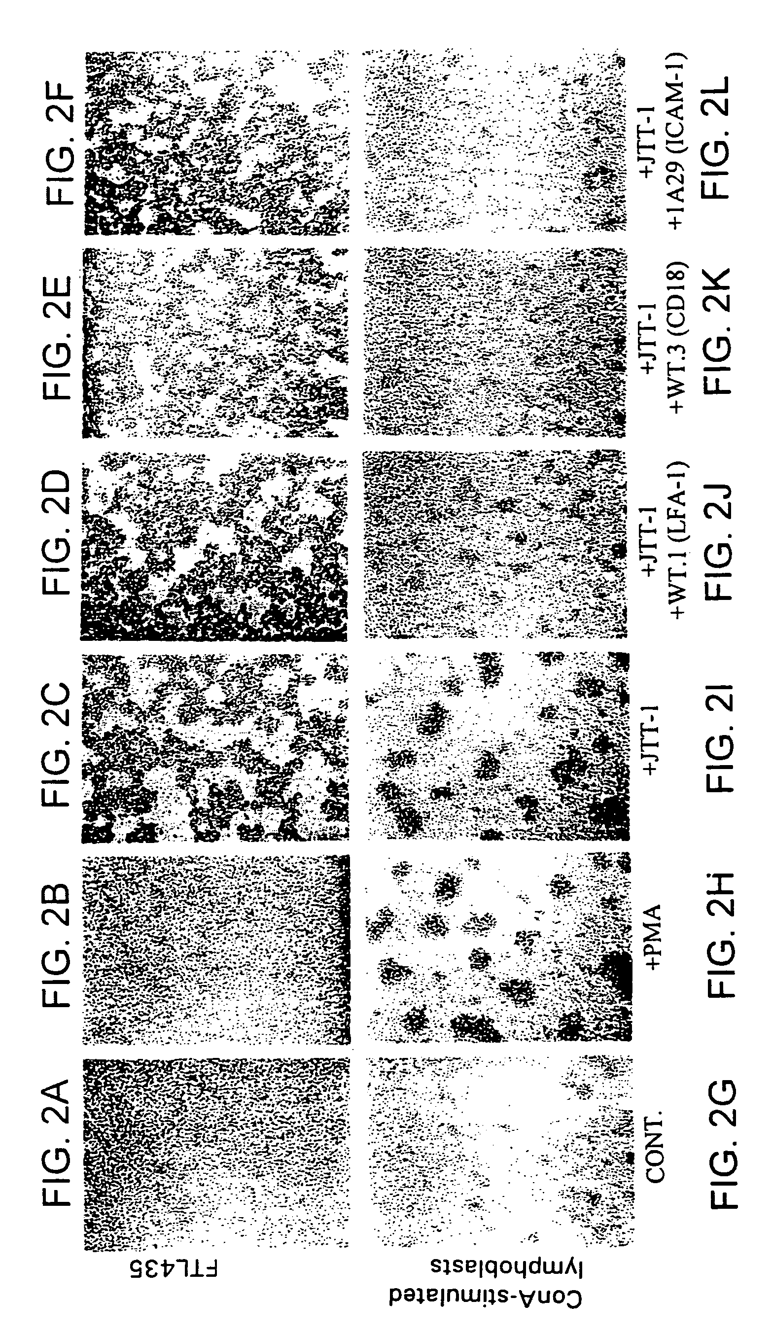
[0338]The hybridomas prepared in Example 1 were screened by analyzing the effect of the antibodies produced in the culture supernatant of the hybridomas on FTL435 cells, which were used as the immunogen. FTL435 cells (5×10⁶ cells / ml, 0.1 ml) were seeded into each well of a 96-well microtiter plate and cultivated at 37° C. for an hour in the presence of culture supernatant of each hybridoma (10 μg / ml each). The results obtained for hybridoma clones “JTT-1” and “JTT-2” are shown in FIG. 1 and FIG. 2.

[0339]It was revealed that a monoclonal antibody produced by hybridoma clone “JTT-1” (“JTT-1 antibody”) strongly agglutinated FTL435 cells (FIG. 1(b) and FIG. 2(c)) and that addition of “JTT-2 antibody” strongly inhibited the aggregation of FTL435 cells induced by “JTT-1 antibody” stimulation (FIG. 1(d)). The assays, in which no hybridoma supernatant was added, were used as controls (FIG. 1(a) and FIG. 2(a)).

[0340]In orde...

example 3

Reactivity of “JTT-1 Antibody” and “JTT-2 Antibody” to Various Cells

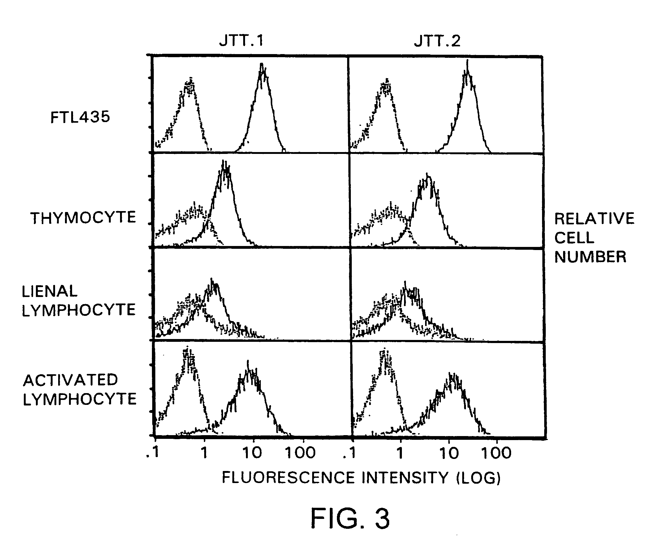
[0347]In order to analyze the expression pattern of molecules recognized by “JTT-1 antibody” and “JTT-2 antibody” in various cells, the reactivities of the antibodies to various cells were examined. Molecules recognized by “JTT-1 antibody” or “JTT-2 antibody” are designated “JTT-1 antigen” or “JTT-2 antigen”, respectively.

[0348]A five- to ten-week-old Wistar rat (150 to 250 g) was killed by anesthesia with diethyl ether. The thymus and spleen were taken out of its chest and abdomen, respectively, by celiotomy, and homogenized to prepare cell suspension. The resulting spleen cells were cultivated in RPMI1640 medium containing 2 μg / ml concanavalin A and 10% FCS at 37° C. for 3 days to prepare activated lymphoblasts.

[0349]FTL435 cells, thymocytes, spleen cells, and activated lymphoblasts (5×10 5 cells each) were reacted with “JTT-1 antibody” or “JTT-2 antibody” and then with FITC-labeled anti-mouse IgG (Cappel). The fl...


PUM

PropertyMeasurementUnit
temperatureaaaaaaaaaa
pHaaaaaaaaaa
pHaaaaaaaaaa

Abstract

Novel cell surface molecules recognized by monoclonal antibodies against a cell surface molecule of lymphocytic cells that play an important role in autoimmune diseases and allergic diseases have been isolated, identified, and analyzed for their functions. The cell surface molecules are expressed specifically in thymocytes, lymphocytes activated by ConA-stimulation, and peripheral blood lymphocytes, and induce cell adhesion. Antibodies against the cell surface molecules significantly ameliorate pathological conditions of autoimmune diseases and allergic diseases.

Description

CROSS REFERENCE TO RELATED APPLICATIONS[0001]This application is a continuation and claims the benefit of priority under 35 USC §120 of U.S. application Ser. No. 09 / 383,551, filed Aug. 26, 1999 now U.S. Pat. No. 7,030,225, which is a continuation-in-part of international application number PCT / JP98 / 00837, filed Feb. 27, 1998, which claims the benefit of priority under 35 USC §119 of Japanese application number 9 / 62290, filed Feb. 27, 1997, and Japanese application number 10 / 62217, filed Feb. 26, 1998. The disclosures of the prior applications are considered part of (and are incorporated by reference in) the disclosure of this application.TECHNICAL FIELD[0002]The present invention relates to novel cell surface molecules of mammals; polypeptides and their fragments constituting the molecules; fusion polypeptides comprising the polypeptide fragments and immunoglobulin fragments; genes encoding the polypeptides and the fragments; vectors comprising the genes; transformants into which th...

Claims


Application Information

Patent Timeline
Patent Type & Authority Patents(United States)
IPC IPC(8): C07K16/00A61K39/395C12N5/12A01K67/027A61K38/00A61P29/00A61P37/00A61P37/02C07K14/47C07K14/705C07K16/18C07K16/28C07K19/00C12N1/21C12N5/00C12N5/10C12N15/02C12N15/09C12N15/12C12P21/02C12P21/08C12R1/19C12R1/91
CPCC07K14/705C07K14/70521C07K14/4713A01K2217/05A01K2217/075A61K38/00C07K2319/00A61P29/00A61P37/00A61P37/02A61P37/06A61P37/08
Inventor TAMATANI, TAKUYATEZUKA, KATSUNARI
Owner JAPAN TOBACCO INC
Why Eureka
Try Eureka