Pluripotent stem cell growing method

a stem cell and growing method technology, applied in the field of growing methods and gene transfer methods for pluripotent stem cells, can solve the problems of low proliferation potency of stem cells, inability to obtain stem cells from living tissues, and inability to culturing methods employed for these classical es/eg cells. to achieve the effect of increasing the proliferation potency of cells and increasing the efficiency of gene transfer

Inactive Publication Date: 2007-07-05
AKAIKE TOSHIHIRO
View PDF0 Cites 99 Cited by
  • Summary
  • Abstract
  • Description
  • Claims
  • Application Information

AI Technical Summary

Benefits of technology

[0038] In light of these circumstances, it is an object of the present invention to provide a method for culturing pluripotent stem cells such a

Problems solved by technology

However, stem cells present in parenchymal organs such as the brain, heart or liver are technically difficult to obtain from living tissues and/or to culture in vitro, and such stem cells also generally have low proliferation potency.
Stem cells can also be recovered from tissues from corpses, but the medical use of cells obtained in this manner is associated with ethical problems.
However, the culturing methods employed for these classical ES/EG cells are not suitable methods when human ES (or EG) cells are used for regenerative medicine or other practical purposes.
The use of feeder cells itself is another problem because as such co-culturing systems increase production cost and are poorly suited for large-scale culturing, while separation and purification of the ES cells from the feeder cells is required when the ES cells are to be actually used.
In the future, when human ES cells and other pluripotent stem cells are utilized as cell sources for regenerative medicine, and particularly for cell transplantation therapy, the use of non-human animal cell products such as MEF cells and FBS will not be desirable because of risks including potential infection of the ES cells by heterozoic viruses and contamination with antigenic molecules that may be recognized as heteroantigens (Martin et al., Nature Med. 11:228, 2005).
Given the active, i.e., proliferating, nature of ES cells in comparison to other cell types, few attempts have been made to actually investigate their proliferation potency; however, the needs of regenerative medicine will require increased proliferation of such cells.
One of the problems currently encountered in culturing pluripotent stem cells is that the cells generally form tight colonies and are therefore difficult to handle for passaging and the like.
When this is done, however, dispersion of the ES/EG cell colonies into individual cells requires relatively high-concentration protease treatment and/or vigorous mechanical stirring, and such procedures significantly reduce the viability and adhesion

Method used


Image

  • Pluripotent stem cell growing method
  • Pluripotent stem cell growing method
  • Pluripotent stem cell growing method

Examples


example 1

Preparation of Recombinant E-Cadherin Protein

[0125] The methods for construction of a vector for expression of a fusion protein comprising the murine E-cadherin extracellular region and IgG Fc portion (IgG / Fc) (hereinafter referred to as “E-cad-Fc”), and for production and purification of the protein were based on methods reported by the present inventors (Nagaoka et al., Biotechnol. Lett. 24:1857, 2002 (Non-patent document 6); Protein Eng. 16:243, 2003 (Non-patent document 7)). First, an extracellular domain (E-cad-ECD)-coding DNA fragment (corresponding to amino acid residues 1-699) from E-cadherin cDNA was amplified using as template cDNA containing the full-length sequence for murine E-cadherin (allotted by RIKEN; RDB No. 1184) as template. A DNA fragment coding for murine IgG / Fc was isolated from cDNA obtained by preparing mRNA from a murine IgG1-expressing hybridoma and performing reverse transcription with Reverse Transcriptase. After confirming the nucleotide sequences of b...

example 2

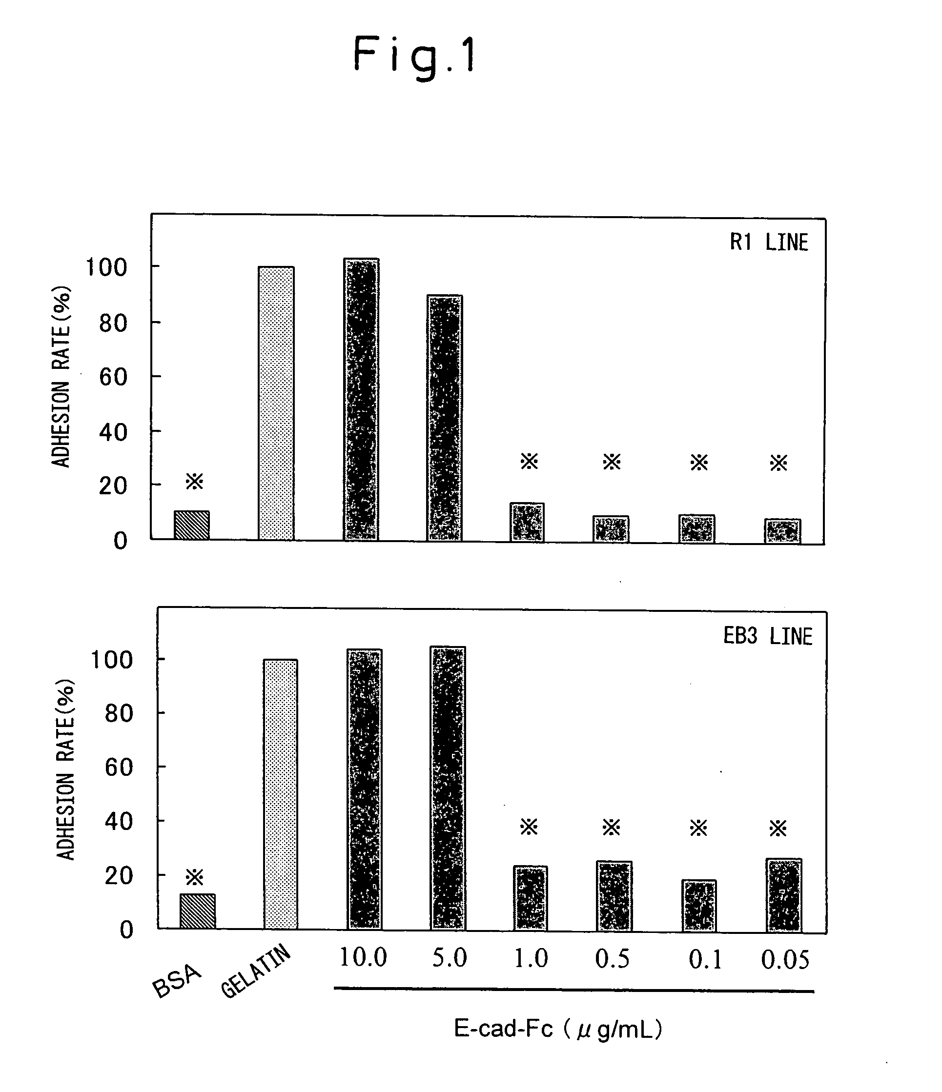
Adhesion of ES Cells to E-Cad-Fc Plate

[0128] The adhesion of ES cells to a cell culturing plate coated with the E-cad-Fc protein (hereunder, “E-cad-Fc plate”) was examined. A PBS-diluted solution of the E-cad-Fc protein was poured into untreated polystyrene culturing plates of different sizes and treated for coating overnight at 37° C. After rinsing and before seeding of the cells, blocking treatment was performed for 1-2 hours with 0.1% BSA solution to prevent non-specific adhesion of the cells. As a control, plates coated with BSA (0.1%), gelatin (0.1%), type I collagen (0.01%; KOKEN) or fibronectin (5.0 μg / mL; KOKEN) were used.

[0129] The ES cell lines used were EB3 cells (provided by Prof. Hitoshi Niwa of RIKEN), R1 cells (Nagy et al., Proc. Natl. Acad. Sci. USA 90:8424, 1993) and 129SV cells (obtained from Dainippon Pharmaceutical Co. Ltd.), and the experimental results showed no differences between the different ES cell lines. These ES cells were passaged according to the met...

example 3

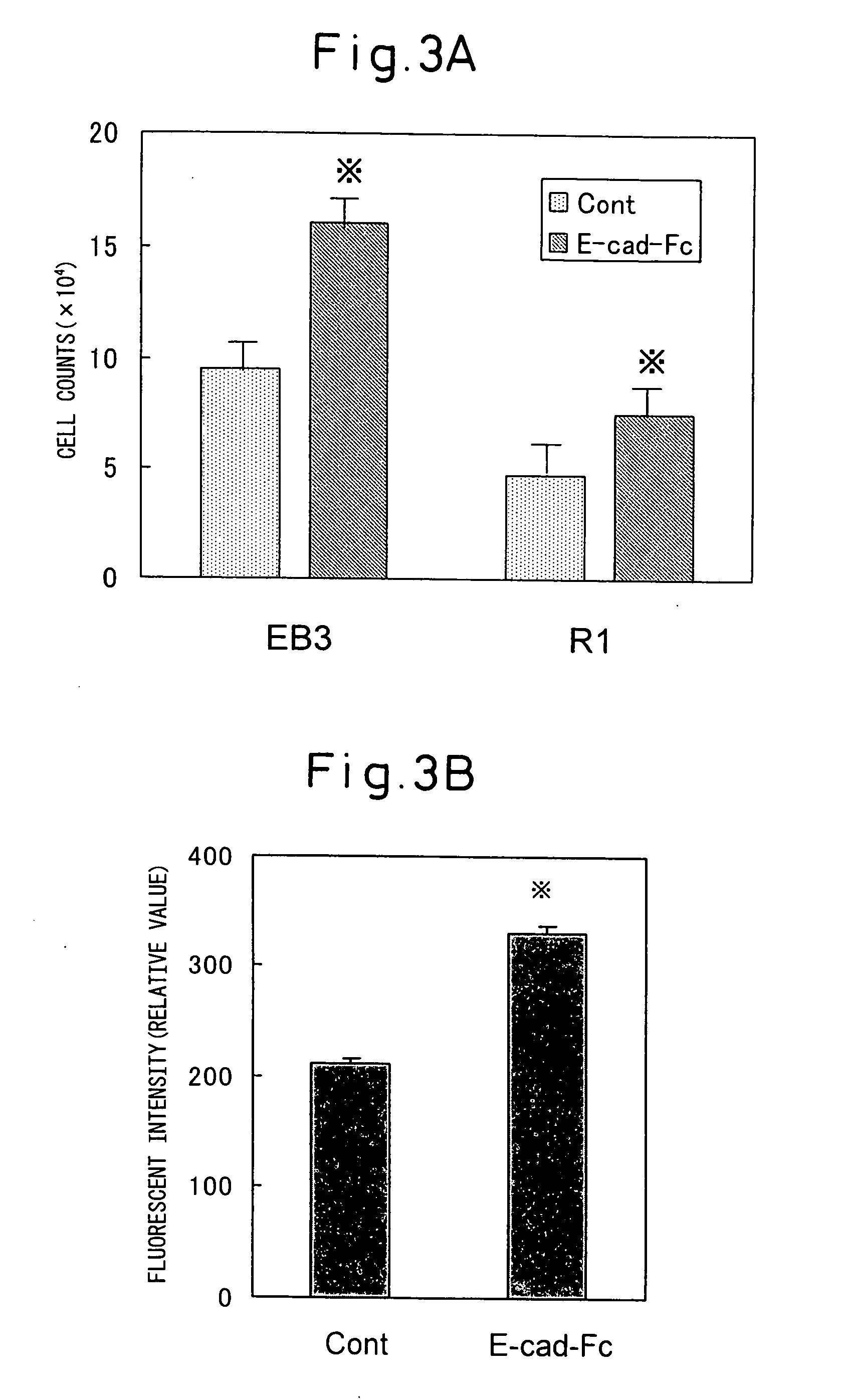
Culturing of ES Cells Using E-Cad-Fc Plate

[0134] In order to examine the proliferation potency of ES cells on an E-cad-Fc plate, ES cells passaged under ordinary conditions were recovered and 500 of the ES cells were seeded on an E-cad-Fc plate or a gelatin plate (96-well plate) and cultured for 3 to 4 days. After rinsing the cells with serum-free medium, the cell counts were measured with Alamar Blue in the same manner as above. As a result, the number of ES cells cultured on the E-cad-Fc plate with respect to the number of ES cells cultured on the gelatin plate by day 3 of culturing was significantly higher for both the EB3 and R1 cell lines (see FIG. 3A). Also, the cell counts of the E-cad-Fc plate groups with both ES cell lines were approximately 2 times greater by day 4 of culturing. When the ES cells were recovered after four similar passages and the counts were recorded, the numbers of E-cad-Fc plate-cultured ES cells were more than 3-5 times greater than those cultured on t...


PUM

PropertyMeasurementUnit
Concentrationaaaaaaaaaa
Adhesivityaaaaaaaaaa

Abstract

A novel growing method is provided for pluripotent stem cells such as ES cells. The method of the invention is a pluripotent stem cell growing method and gene transfer method in which pluripotent stem cells are cultured under conditions that maintain their undifferentiated state and pluripotency, the method being characterized by using a liquid medium and a culturing vessel having immobilized or coated on a substrate solid phase surface a molecule which is adhesive to the pluripotent stem cells in a fixed concentration, to grow the pluripotent stem cells in a dispersed state while maintaining their undifferentiated state and pluripotency, without using feeder cells, or to transfer and express a gene therein.

Description

TECHNICAL FIELD [0001] The present invention relates to a growing method and to a gene transfer method for pluripotent stem cells such as ES cells, and to pluripotent stem cells prepared by the methods. BACKGROUND ART [0002] In order to continue to live, organisms have the ability to rapidly replace and repair lost or damaged cells and tissue, and this ability is known as “regenerative capacity”. Examples of “regenerative capacity” in higher animals include the commonly known phenomena of wound healing of skin and blood vessels, but even parenchymal organs such as the liver and kidneys are known to undergo cell growth and tissue reconstruction for rapid restoration of tissue homeostasis in response to tissue damage. Recent years have seen attempts to utilize this innate “regenerative capacity” of biological organisms to achieve cures or amelioration of various diseases and wounds, and such new medical techniques are coming to be known as “regenerative medicine”. [0003] Stem cells pl...

Claims


Application Information

Patent Timeline
IPC IPC(8): C12N15/09C12N5/06C12N5/08C12N5/0789C07K14/47C12N5/071
CPCC12N2533/50C12N5/0606C07K14/435C07K14/47C12N5/06C12N5/0602
Inventor AKAIKE, TOSHIHIROFUKUDANAGAOKA, MASATOKOSHIMIZU, UICHI
Owner AKAIKE TOSHIHIRO
Why Eureka
Try Eureka