Optical contrast agents for optically modifying incident radiation

a technology of incident radiation and optical attenuation, which is applied in the field of optical contrast agents, can solve the problems of limited image depth, insufficient emulsification, and insufficient production of long-lived microparticles, and achieve the effect of enhancing image contras

Active Publication Date: 2007-04-03
THE BOARD OF TRUSTEES OF THE UNIV OF ILLINOIS
View PDF34 Cites 22 Cited by
  • Summary
  • Abstract
  • Description
  • Claims
  • Application Information

AI Technical Summary

Benefits of technology

[0016]In a third aspect, the present invention is a method of forming an image by optical coherence tomography, including exposing a patient to electromagnetic radiation, collecting reflected electromagnetic radiation, and forming an image from the collected electromagnetic radiation, the improvement comprising administering a contrast agent to the patient to enhance contrast of the image.

Problems solved by technology

In tissues other than the eye, the imaging depth is limited by optical attenuation due to scattering and absorption.
Alone, however, emulsification is insufficient to produce long-lived microparticles.

Method used


Image

  • Optical contrast agents for optically modifying incident radiation
  • Optical contrast agents for optically modifying incident radiation
  • Optical contrast agents for optically modifying incident radiation

Examples


examples

[0082]The examples herein are illustrations of various embodiments of this invention and are not intended to limit it in any way.

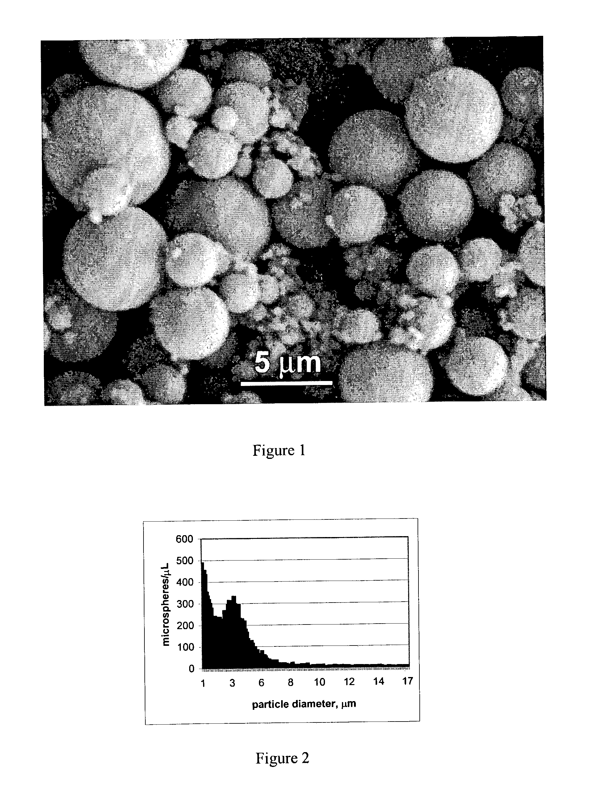
[0083]The contrast agents investigated in this study are similar to those used in ultrasound echocardiography [4]. These agents are hollow microparticles 0.5 to 5 microns in diameter with a 50 Å thick protein shell. FIG. 1 shows a scanning electron micrograph of the microparticles. The microparticles utilized were air-filled and produced by sonicating a 5% weight per volume solution of bovine serum albumin (BSA) in water. The high-intensity ultrasound necessary for the reaction was generated by a titanium horn with tip diameter of 0.5 inches, driven at 20 kHz. The BSA solution was sonicated for 3 minutes at an acoustic power of 76 W / cm2 [3]. The microparticles may be re-suspended with 0.1 M 4-morpholine ethane sulfonic acid, pH=4.5. The diameter of the microparticles is dependent on the acoustic power and the frequency of ultrasound used. Diameters ranging...


PUM

PropertyMeasurementUnit
wavelengthaaaaaaaaaa
depthsaaaaaaaaaa
frequencyaaaaaaaaaa

Abstract

A method of enhancing the contrast of an image of a sample, comprises forming an image of a mixture, by exposing the mixture to electromagnetic radiation. The mixture comprises the sample and microparticles. The enhancement is particularly suitable for optical coherence tomography.

Description

FEDERALLY SPONSORED RESEARCH OR DEVELOPMENT[0001]The subject matter of this application may in part have been funded by the National Science Foundation (BES-0086696). The government may have certain rights in this invention.BACKGROUND[0002]When imaging biological tissues, it is often desirable to enhance the signals measured from specific structures. Contrast agents, which produce a strong emission or reflection signal, have been utilized in virtually every imaging modality including ultrasound [2], computed tomography [57], magnetic resonance imaging [58], and optical microscopy [59].[0003]Optical coherence tomography (OCT) is an emerging high-resolution medical and biological imaging technology [15–21]. OCT is analogous to ultrasound B-mode imaging except reflections of low-coherence light are detected rather than sound. OCT detects changes in the backscattered amplitude and phase of light.[0004]Cross-sectional OCT imaging is performed by measuring the backscattered intensity of l...

Claims


Application Information

Patent Timeline
Patent Type & Authority Patents(United States)
IPC IPC(8): A61K49/00A61K9/16A61K9/50A61K47/48
CPCA61K9/1694A61K9/5089A61K47/48869A61K47/6925
Inventor BOPPART, STEPHEN A.MARKS, DANIEL L.SUSLICK, KENNETH S.TOUBLAN, FARAH JEAN-JACQUES
Owner THE BOARD OF TRUSTEES OF THE UNIV OF ILLINOIS
Why Eureka
Try Eureka