[0007]In accordance with one aspect of the invention, the invention is directed to a device and / or method for visually combining patient image data from
transillumination and / or tomographic imaging methods and / or object image data comprising video images, which overcome the disadvantages of the prior art. The device and / or method can enable desired image information to be received directly and in the direct view of the patient and thus to also exercise an influence on assisting the treatment.
[0009]This enables the physician carrying out the treatment to hold the image
display device himself, with the associated camera, in front of the part of the patient's body and to read off the desired anatomical information while looking directly at the patient. Just by associating the video camera and image display device and forming them as a portable unit, the device in accordance with the invention takes another step towards creating a “glass patient”, the image display device merely having to be held in front of the part of the patient's body in question. Since the association of the camera to the image display device is tracked via the tracking means, the position of the elements viewed is always known, and the navigation
system has information on how and from which direction the part of the patient's body is being viewed. For example, when performing minimally
invasive surgery, in which no area operated on is exposed, the device in accordance with the invention thus offers the physician carrying out the treatment the option, during the operation, of exactly apprizing himself once again of the position of the patient's anatomical parts and / or of other objects in relation to the patient's
anatomy. Tracked and / or navigated instruments can also be displayed on the image display device. The physician no longer has to look away from the patient in order to look into the interior of the patient's body.
[0010]In addition, the physician is put into a position to make changes and modifications which assist his work, via the
input device, while still directly viewing the patient and with the aid of information from the image display. He can then verify the effect of his modifications directly and while viewing the patient without having to firstly turn toward an input
station, which makes verifying or correcting particular measures much simpler and more direct.
[0017]In accordance with another embodiment, changes to the patient body predicted by planning are displayed on the image display device by inputs at the
input device. This enables planning to be directly controlled while directly viewing the part of the patient's body.
[0022]The image display device can be designed to be light and easy to
handle, for example, by designing it as a portable flat-screen, such as an LCD flat-screen. As noted above, this screen can of course exhibit the touch screen function. The device in accordance with the invention can be supplied with power either by a power supply of its own (a battery or power pack) or via cables. If
wireless, such as radio, transmission is used for the data, then a power supply of its own is particularly suitable for the device, in order to achieve the greatest freedom of handling.
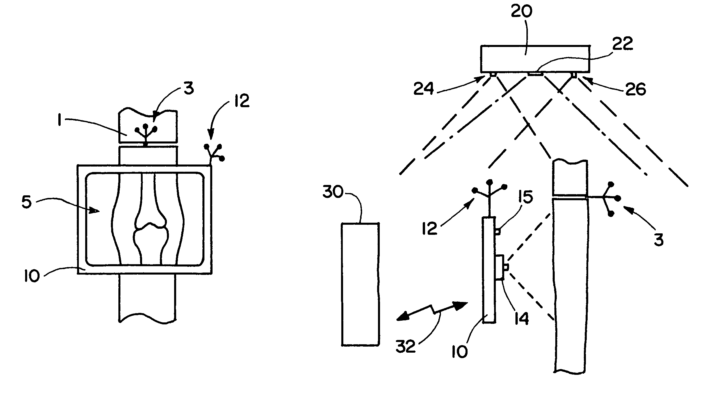
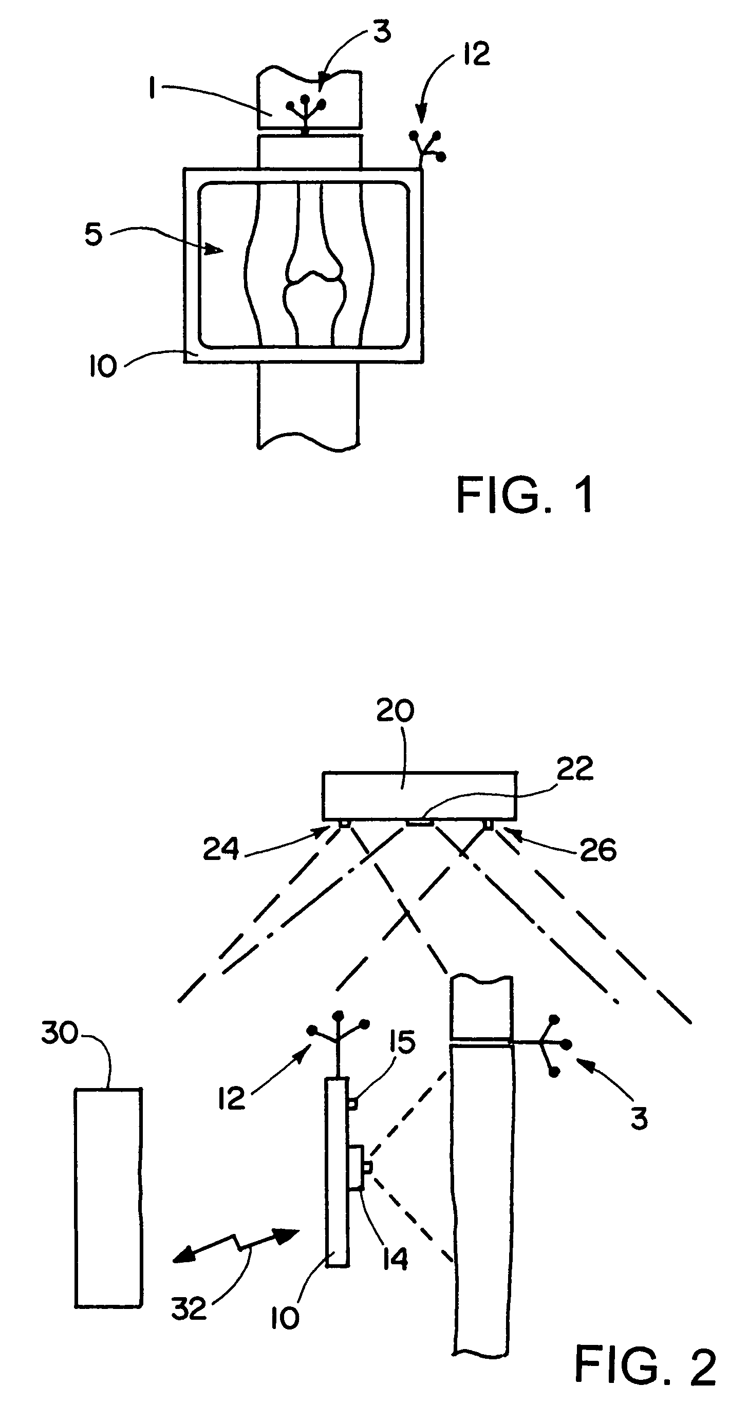
[0023]The video camera can exhibit a small aperture and a low
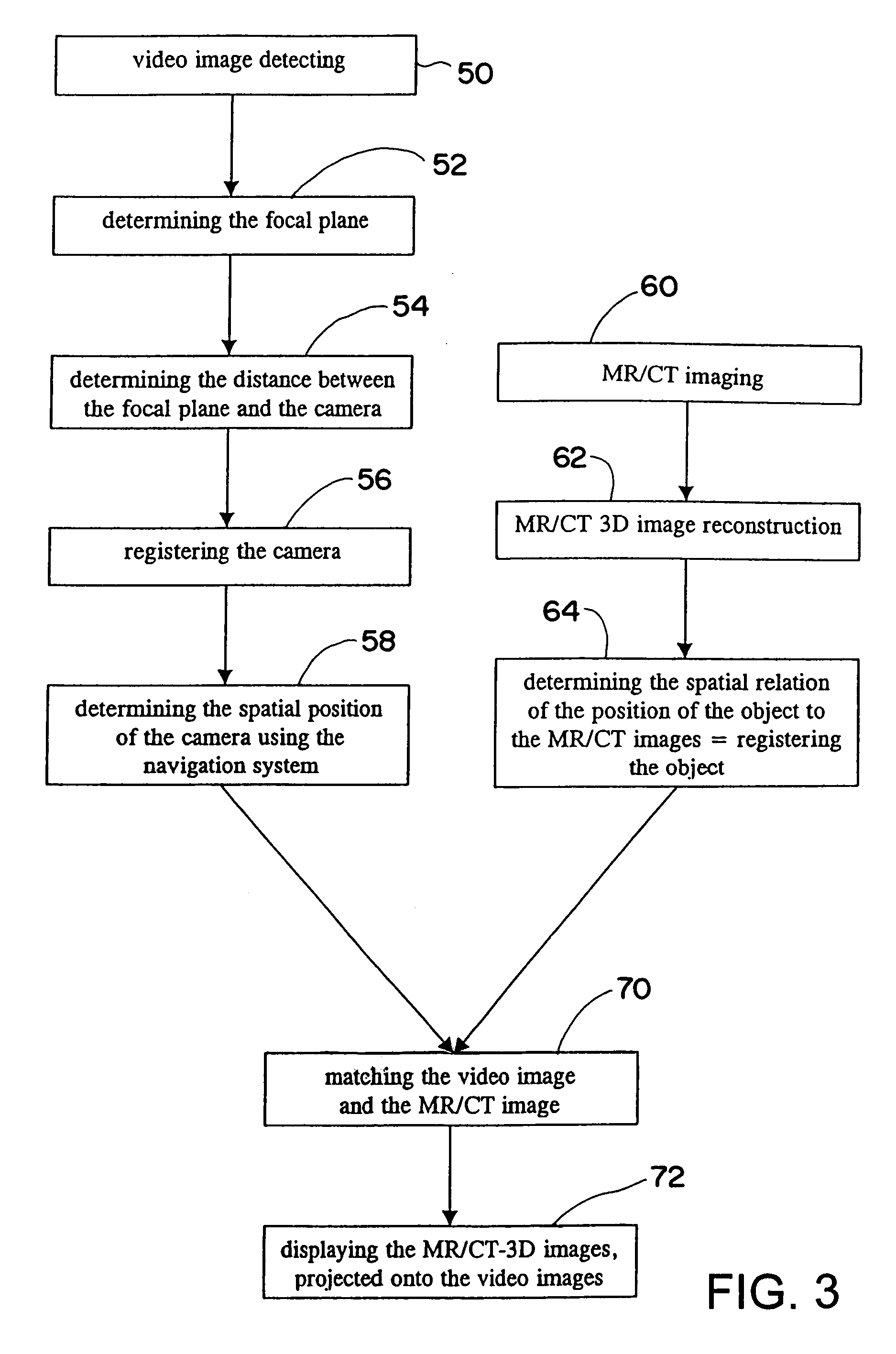
depth of field, so that only a small area of the captured image is in the focal plane. Using such a camera, it is possible ascertain the distance between the image and the focal plane. If the spatial position of the camera is known from the navigation
system, and the distance from the
image plane is likewise known, then the computer system connected to the navigation system can calculate the spatial position of the
video image in real time. It is therefore possible to optimally associate the
video image and the image data from previous
transillumination and / or tomographic imaging, or equally to associate object data.

