Apparatus and method for registering 2D radiographic images with images reconstructed from 3D scan data

a radiographic image and scan data technology, applied in the field of two-dimensional radiographic images with scan data reconstructed from 3d (three-dimensional) scan data, can solve the problems of inability to achieve intensity-based methods, inability to perform radiosurgery, and inability to achieve long computation time, so as to achieve precise and rapid results and improve accuracy

Inactive Publication Date: 2007-04-17
ACCURAY
View PDF5 Cites 188 Cited by
  • Summary
  • Abstract
  • Description
  • Claims
  • Application Information

AI Technical Summary

Benefits of technology

[0009]The present invention is directed to a precise and rapid method and system for registering 2D x-ray images with images that have been reconstructed from 3D scan data. A hierarchical and iterative framework for the registration algorithm permits a higher accuracy to be achieved, in both the translational and rotational adjustments. The total computation time is about an order of magnitude faster than other techniques existing in the prior art.
[0012]In order to determine these parameters, a 3D multi-level matching is first performed, in order to determine an initial estimate for the in-plane transformation parameters (x, y, θ). Based on these parameters (x, y, θ) obtained by 3D multi-level matching, an initial 1-D search is performed for each of the pair of out-of-plane rotation parameters (r, Φ). The in-plane translation parameters (x, y) are then refined, using 2D sub-pixel matching, to increase the accuracy of these parameters.

Problems solved by technology

Because the full information content of the image is not used, however, accuracy is sacrificed.
Because a lengthy computation time is required, however, intensity-based methods are not practical for purposes of radiosurgery, or for clinical practice in general.
Unfortunately, it is difficult to meet both requirements simultaneously, because of several reasons.
Establishing this relationship is difficult, so a linear attenuation model is often used.
However, the skeletal structures in DRR images cannot be reconstructed very well using the linear model, the DRRs being only synthetic x-ray projection images.
Full resolution images are rarely used, however, due to the extremely slow computation that results from using such images.
However, the accuracy and stability of the estimates are not sufficient to meet the sub-mm precision that is required in radiosurgery applications.

Method used


Image

  • Apparatus and method for registering 2D radiographic images with images reconstructed from 3D scan data
  • Apparatus and method for registering 2D radiographic images with images reconstructed from 3D scan data
  • Apparatus and method for registering 2D radiographic images with images reconstructed from 3D scan data

Examples


Embodiment Construction

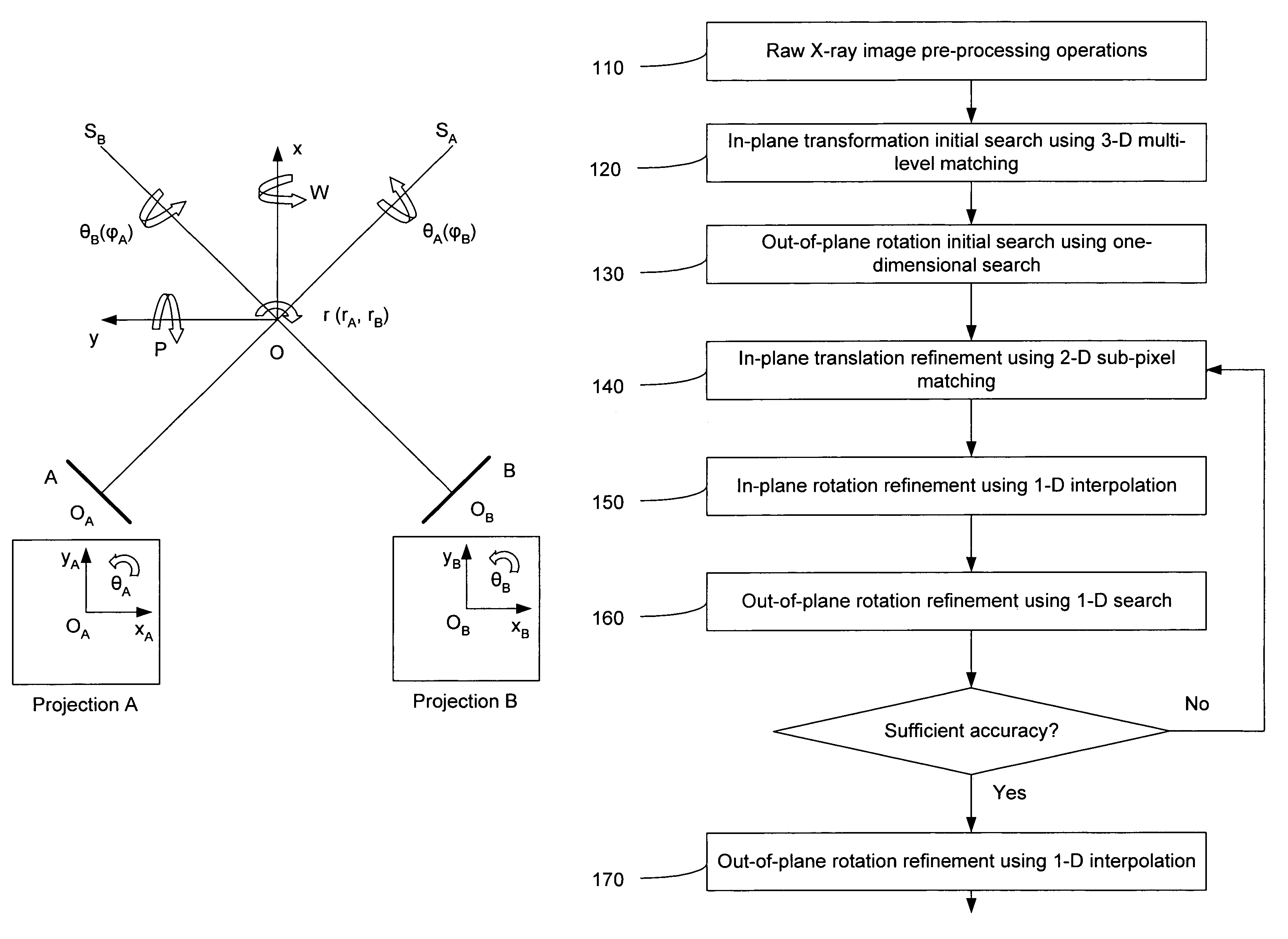
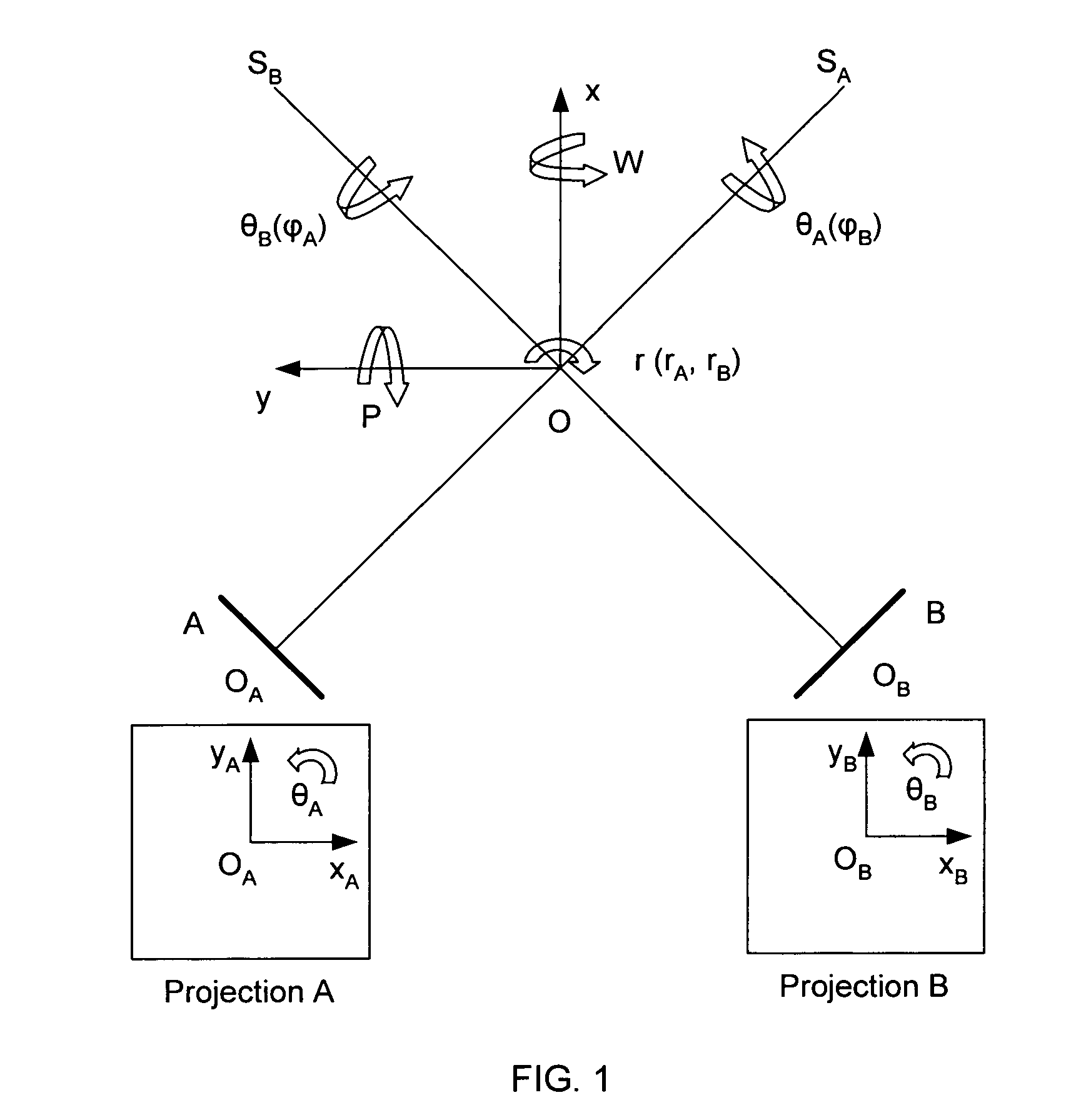
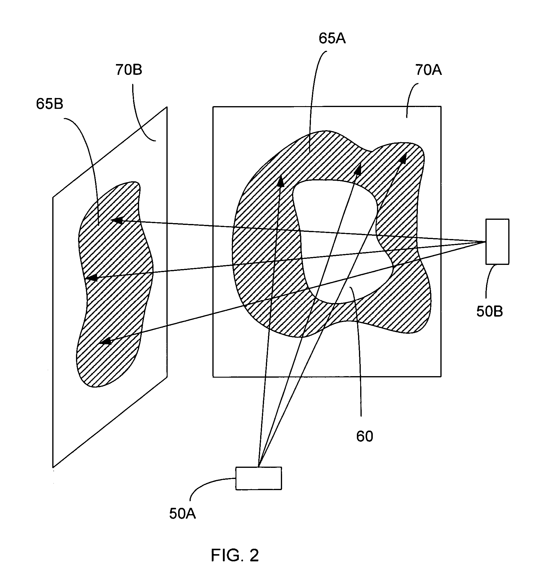
[0020]The present invention is directed to an improved method and system for performing medical 2D / 3D registration. The tracking method and system of the present invention is useful in radiosurgery and radiotherapy; however the method and system of the present invention can also be used in applications other than radiosurgery and radiotherapy, i.e. in any application where there a need to track rigid object by registering 2D radiographic images onto 3D scan data. While 2D x-ray images are described in the preferred embodiment of the present invention, the present invention can be used for the registration of any other type of 2D images onto 3D scan data.

[0021]In overview, the method of the present invention includes generating a reconstructed 2D image from the 3D scan data. The change in the position of the target (or other rigid object) in the radiographic image, as compared to the position of the target in the 3D scan data (as indicated in the reconstructed 2D image) is described ...


PUM


Abstract

A method and system is provided for registering a 2D radiographic image of a target with previously generated 3D scan data of the target. A reconstructed 2D image is generated from the 3D scan data. The radiographic 2D image is registered with the reconstructed 2D images to determine the values of in-plane transformation parameters (x, y, θ) and out-of-plane rotational parameters (r, Φ), where the parameters represent the difference in the position of the target in the radiographic image, as compared to the 2D reconstructed image. An initial estimate for the in-plane transformation parameters is made by a 3D multi-level matching process, using the sum-of-square differences similarity measure. Based on these estimated parameters, an initial 1-D search is performed for the out-of-plane rotation parameters (r, Φ), using a pattern intensity similarity measure. The in-plane parameters (x, y, θ) and out-of-plane parameters (r, Φ) are iteratively refined, until a desired accuracy is reached.

Description

FIELD OF THE INVENTION[0001]The present invention relates to a method and system for registering 2D (two-dimensional) radiographic images with images reconstructed from 3D (three-dimensional) scan data. More particularly, the invention relates to a method and system for registering 2D stereo x-ray image data with digitally reconstructed radiographs of 3D CT scan data.BACKGROUND[0002]Medical image registration is useful in many areas of medicine, for example radiosurgery. In radiosurgery, tumors and other lesions are treated by delivering a prescribed high dose of high-energy radiation to the target area, while minimizing radiation exposure to the surrounding tissue. Radiosurgery therefore calls for an ability to accurately focus on a target region, so that only the target receives the desired high doses of radiation, while surrounding critical structures are avoided. Typically, 3D imaging modalities, such as computed tomography (CT), magnetic resonance (MR) imaging, or positron emis...

Claims


Application Information

Patent Timeline
Patent Type & Authority Patents(United States)
IPC IPC(8): A61N5/10G01N23/04G06T7/00G06T7/20
CPCA61N5/1049G01N23/04G06T7/0026G06T7/0038G06T7/2053A61N5/1067A61N5/107A61N2005/1061A61N2005/1062G06T2207/10124G06T2207/30004G06T7/32G06T7/38G06T7/254
Inventor FU, DONGSHANKUDUVALLI, GOPINATHQURESHI, SHEHRZAD
Owner ACCURAY
Why Eureka
Try Eureka