Method for determining the efficacy of an anti-cancer treatment using image analysis

a technology of image analysis and anti-cancer treatment, applied in the field of ultrasonic image analysis, can solve the problems of inflammatory response, rapid deterioration, labor intensive, etc., and achieve the effects of reducing error variance, expanding statistical inference, and increasing discrimination between regions

Inactive Publication Date: 2007-06-12
PFIZER INC
View PDF7 Cites 22 Cited by
  • Summary
  • Abstract
  • Description
  • Claims
  • Application Information

AI Technical Summary

Benefits of technology

[0026]These objectives are accomplished by the use of powerful multivariate statistical analysis methods that take advantage of the joint probability distribution function to increase both specificity and sensitivity of the image segmentation. In addition to the immediate advantages to the specific application to cancer research, this method may be adapted to the segmentation of other tissue regions of interest. The experimental results and the analysis of images obtained by the apparatus and method of the invention show that the multivariate analysis of ultrasound images more accurately characterize tumors by properly segmenting viable from necrotic tissue.
[0039]The above capability allows for a more detailed in vivo analysis of tumor growth beyond simple dimensional measurements and may be useful in the analysis of tumor angiogenesis biomarkers from Power Doppler. Consequently, longitudinal studies can be done that take advantage of the within-subjects correlation of repeated measurements, reducing the error variance, expanding the statistical inference to the population of subjects and increasing the power of the analysis.

Problems solved by technology

This conventional approach necessarily requires more specimens (i.e. animals) than a longitudinal experiment over time, and is very labor intensive.
Cells, upon death, elicit an inflammatory response and quickly deteriorate.
However, histopathology is necessarily post-mortem, and a longitudinal analysis of the tumor over time is not possible.
In laboratory research, where the number of animals used for research can be quite large, histopathology measurements are time consuming, expensive and require terminating the study of the subject.
Additionally, tissue fixing can result in damage to some aspect of the tumor and loss of information.
This process is time consuming, requires the use of a large number of subjects, and necessarily precludes the use of longitudinal measurements on the same tumor.
In addition to the statistical problems posed by the lack of repeated measurements, the histological analysis of tumors is done using microscopically thin slices of the tumor, providing information on a very limited region, in a non-realtime manner.
In the late 1980's and early 1990's, ultrasonic tissue characterization had mixed results due to the characteristic coherent interference phenomenon known as “speckle”.
However, tumor cells, like all living cells, can die if exposed to a fatal environment.
However, such high frequency ultrasound devices are relatively expensive, and are not readily available in clinical settings, in comparison to lower frequency devices, e.g., devices operating in the 4–12 MHz region, for example.
Unfortunately, and as determined by the use of histopathologic sampling and training images, this univariate approach results in a higher probability of misclassifying a significant portion of the various pixels into the wrong class.
Further, univariate statistical analysis of image pixel data increases the probability of both Type I and Type II errors, along with the misclassification of pixels.

Method used


Image

  • Method for determining the efficacy of an anti-cancer treatment using image analysis
  • Method for determining the efficacy of an anti-cancer treatment using image analysis
  • Method for determining the efficacy of an anti-cancer treatment using image analysis

Examples


Embodiment Construction

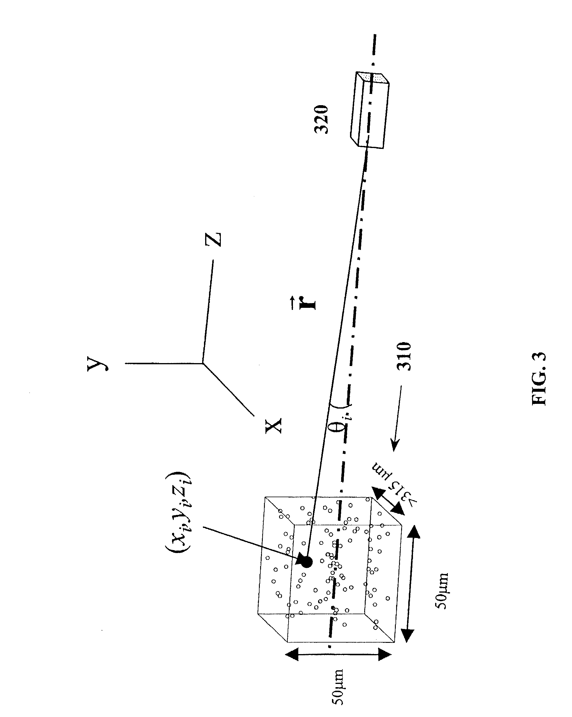
[0059]A first aspect of the invention will be explained with reference to FIGS. 3 and 4, which show the basic geometry of a volume of scatterers corresponding to an image pixel, and a generalized ultrasound system.

[0060]The geometry of a single voxel 310 of scatterers, corresponding to a single pixel within the image, is depicted in FIG. 3. One tissue parameter that can change the brightness of the image is the number of scatterers within a voxel that corresponds to a single pixel. The mean pixel energy is proportional to the number of scatterers, and is related to the energy transmitted by transducer 320. Therefore, if the density of the scattering bodies increases, the number of scatterers within a voxel increases and the pixel brightness increases. Additionally, if the reflectivity of the scattering bodies increases, the pixel energy increases. Further, if the variation in the characteristic impedances increases, then the reflectivity of the region increases and it follows that t...


PUM


Abstract

An apparatus, system, method, and computer readable medium containing computer-executable code for implementing image analysis uses multivariate statistical analysis of sample images, and allows segmentation of the image into different groups or classes, depending on a correlation to one or more sample textures, or sample surface features. In one embodiment, the invention performs multivariate statistical analysis of ultrasound images, wherein a tumor may be characterized by segmenting viable tissue from necrotic tissue, allowing for more detailed in vivo analysis of tumor growth beyond simple dimensional measurements or univariate statistical analysis. Application of the apparatus and method may also be used for characterizing other types of samples having textured features including, for example, tumor angiogenesis biomarkers from Power Doppler.

Description

CROSS-REFERENCE TO RELATED APPLICATIONS[0001]This application claims priority to provisional application No. 60 / 381,064 filed on May 17, 2002 by David L. Raunig, entitled “Apparatus and Method for Statistical Image Analysis”, the entire contents of which are incorporated herein by reference, and for which benefit is claimed under 35 U.S.C. §119(e).FIELD OF THE INVENTION[0002]This invention relates generally to an apparatus, a system, and method for the analysis of images, and more particularly to a method for the analysis of ultrasonic images. This invention further relates even more particularly to an apparatus and method for the analysis of ultrasonic images using multivariate detection techniques, which, in one exemplary embodiment, may be used to differentiate image pixels representing necrotic material from image pixels representing viable tissue within a specified region of interest, and which, in a further aspect of the invention, may be used in a method for treating cancer.B...

Claims


Application Information

Patent Timeline
Patent Type & Authority Patents(United States)
IPC IPC(8): G06K9/00G06T5/00G06T7/00G06T7/40
CPCG06K9/0014G06T7/0012G06T7/0081G06T7/0083G06T7/0087G06T7/404G06T7/606G06T2207/10056G06T2207/10132G06T2207/20104G06T2207/20116G06T2207/30096G06T7/11G06T7/12G06T7/143G06T7/45G06T7/66G06V20/695
Inventor RAUNIG, DAVID L.
Owner PFIZER INC
Why Eureka
Try Eureka