
Mouse model for psoriasis and psoriatic arthritis

a mouse model and psoriatic arthritis technology, applied in the field of psoriasis, can solve the problems of severely hampered research into the pathogenesis of this common skin disorder, and the efforts to transgeneically deliver inflammatory mediators or keratinocyte growth factors to the skin have not completely reproduced the psoriatic phenotyp

Active Publication Date: 2007-07-24
BOEHRINGER INGELHEIM INT GMBH
View PDF5 Cites 1 Cited by
  • Summary
  • Abstract
  • Description
  • Claims
  • Application Information

AI Technical Summary

Benefits of technology

The invention provides an animal model for psoriasis and psoriatic arthritis using a mutant mouse deficient for c-Jun and JunB in the epidermis. The mutant mouse can be produced by crossing a transgenic mouse with a genetic construct containing the c-Jun and JunB genes flanked with recognition sites for a site-specific recombinase with a transgenic mouse expressing a constitutively active or inducible recombinase in the epidermis. The mutant mouse can also be specifically induced by applying an anti-estrogen. The invention also includes a method for testing drugs to treat psoriasis and psoriatic arthritis in the mutant mouse.

Problems solved by technology

However, extensive efforts aimed at transgenically delivered inflammatory mediators or keratinocyte growth factors to the skin have not completely reproduced the psoriatic phenotype (Xia et al., 2003), which has thus far only been faithfully modeled in animals by transplanting psoriatic skin onto mice with severe combined immunodeficiency disease (SCID).
Since there is no naturally occurring animal skin disease mirroring both phenotype and immunopathogenesis of psoriasis, research into the pathogenesis of this common skin disorder has been severely hampered.

Method used


Image

  • Mouse model for psoriasis and psoriatic arthritis
  • Mouse model for psoriasis and psoriatic arthritis
  • Mouse model for psoriasis and psoriatic arthritis

Examples


example 1

Generation of c-junΔep*; junBΔep* Mice

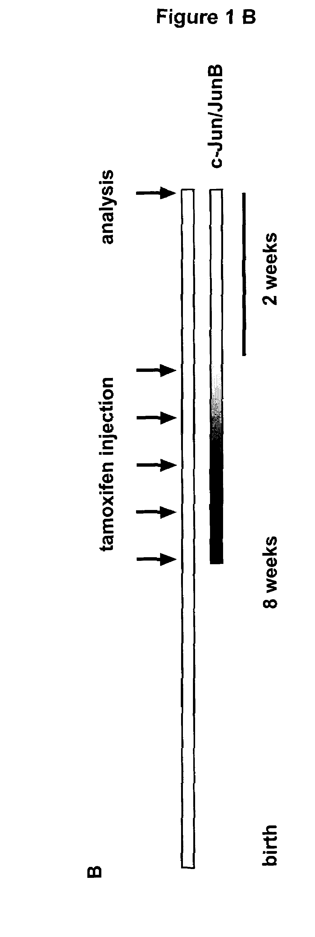
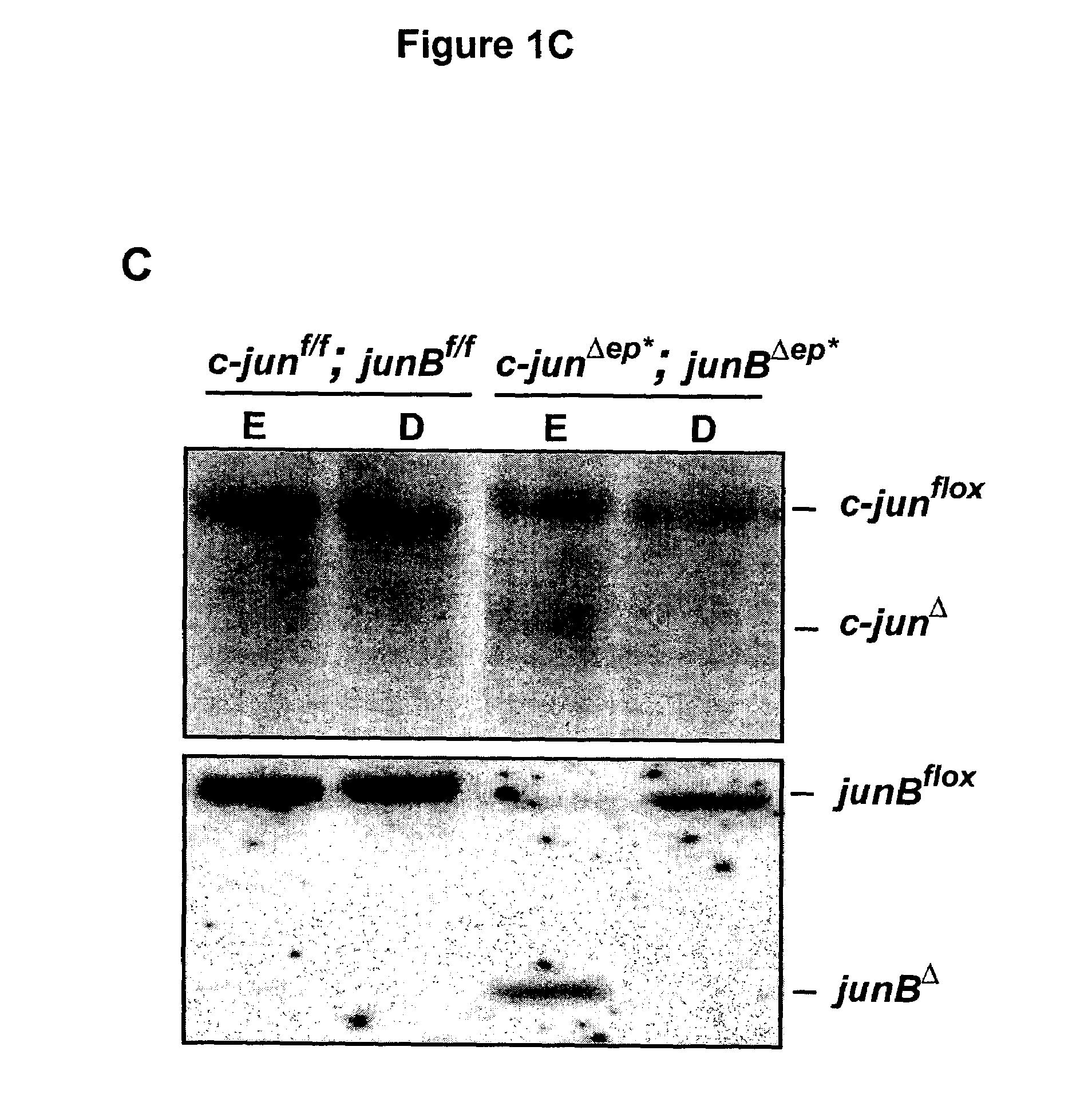
[0072]c-junf / f; junBf / f mice were crossed to K5-Cre-ER transgenic mice and heterozygous progenies were intercrossed to get c-junf / f; junBf / f; K5-Cre-ER and c-junf / f; junBf / f mice. The approach to delete c-jun and junB in the epidermis is outlined in FIGS. 1A and 1B. In brief, 8 weeks old experimental mice (c-junf / f; junBf / f; K5-Cre-ER) and control mice (c-junf / f; junBf / f) were injected intraperitoneally 5 times with 1 mg tamoxifen. 2 weeks after the last injection the mice were analyzed. The deletion of c-jun and junB was analyzed by Southern blot and RPA (RNAse protection assay). Southern blot analyses for c-jun and JunB deletion (FIG. 1C) showed deletion in both cases. The remaining signal for floxed c-jun and floxed junB is explained by incomplete deletion and the inflammatory infiltrate seen in the epidermis of c-junΔep*; junBΔep* mice (FIG. B). Quantification of AP-1 mRNA in the epidermis of c-junΔep*; junBΔep* mice showed significant down ...

example 2

Characterization of c-junΔep*; junBΔep* Mice

[0073]Mice obtained according to the treatment described in Example 1 develop a phenotype resembling psoriasis within 2 weeks. Hairless skin like ear, tail and feet were dramatically affected (FIG. 2A). Histological examination (FIG. 2B) revealed abnormally thickened epidermis, parakeratosis (nucleated keratinocytes in the cornified layer), thickened keratinized upper layer (hyperkeratosis), and fingerlike epidermal projections into the dermis (rete ridges). Furthermore, epidermal microabscesses and the typical inflammatory cell infiltrate are seen: intraepidermal T-cells (CD3 staining), increased numbers of neutrophils in the epidermis (esterase staining), and macrophages in the dermis (esterase- and F4 / 80-staining). H&E staining from affected mouse finger demonstrating granulocytic infiltrates into the joint region resembling psoriatic arthritis (FIG. 2C).


PUM

PropertyMeasurementUnit
morphologyaaaaaaaaaa
conformationalaaaaaaaaaa
protein structureaaaaaaaaaa

Abstract

A mouse that is deficient for c-Jun and JunB in the epidermis or in which deletion of c-Jun and JunB can be specifically induced in the epidermis, and methods for obtaining such mice. The mice and keratinocytes derived therefrom are useful as an animal model for psoriasis and psoriatic arthritis.

Description

[0001]This patent application claims priority to provisional patent application No. 60 / 548,426 filed Feb. 27, 2004, which is hereby incorporated herein by reference.BACKGROUND OF THE INVENTION[0002]1. Field of the Invention[0003]The invention relates to the field of psoriasis.[0004]2. Background Art[0005]Psoriasis, affecting about 2% of the population, is one of the most common human skin disorders that affect skin and joints. It is characterized by complex alterations of various cell types. This includes epidermal hyperproliferation and altered differentiation, as well as angiogenesis and dilation of dermal blood vessels (Schön, 1999; Lebwohl, 2003). In addition, a mixed leukocytic infiltrate is seen. It is composed of activated T lymphocytes, neutrophiles within the dermis and epidermal microabscesses, lining macrophages, and an increased number of dermal mast cells. Cytokines including tumor necrosis factor-α (TNF-α), and interleukin-1 (IL-1), interferon-γ (INF-γ), IL-6, IL-8, va...

Claims


Application Information

Patent Timeline
Patent Type & Authority Patents(United States)
IPC IPC(8): A01K67/027G01N33/00C12N15/85
CPCA01K67/0275C12N15/8509A01K2217/05A01K2227/105A01K2267/03
Inventor ZENZ, RAINERWAGNER, ERWIN
Owner BOEHRINGER INGELHEIM INT GMBH
Learn More