X-ray image-assisted navigation using original, two-dimensional x-ray images

Active Publication Date: 2007-07-31
BRAINLAB +1
View PDF9 Cites 54 Cited by
  • Summary
  • Abstract
  • Description
  • Claims
  • Application Information

AI Technical Summary

Benefits of technology

[0009]It is an object of the present invention to provide a method for x-ray image-assisted navigation, which overcomes the aforementioned disadvantages of the prior art. In particular, the intention is to ensure that the

Problems solved by technology

However in the methods currently used, it has the disadvantage that every image has to be registered individually and then displayed in correct relation to the instruments or apparatus.
While these x-ray images, in their original state, actually provide very good imaging data with a high contrast and good ac

Method used


Image

  • X-ray image-assisted navigation using original, two-dimensional x-ray images
  • X-ray image-assisted navigation using original, two-dimensional x-ray images
  • X-ray image-assisted navigation using original, two-dimensional x-ray images

Examples


Embodiment Construction

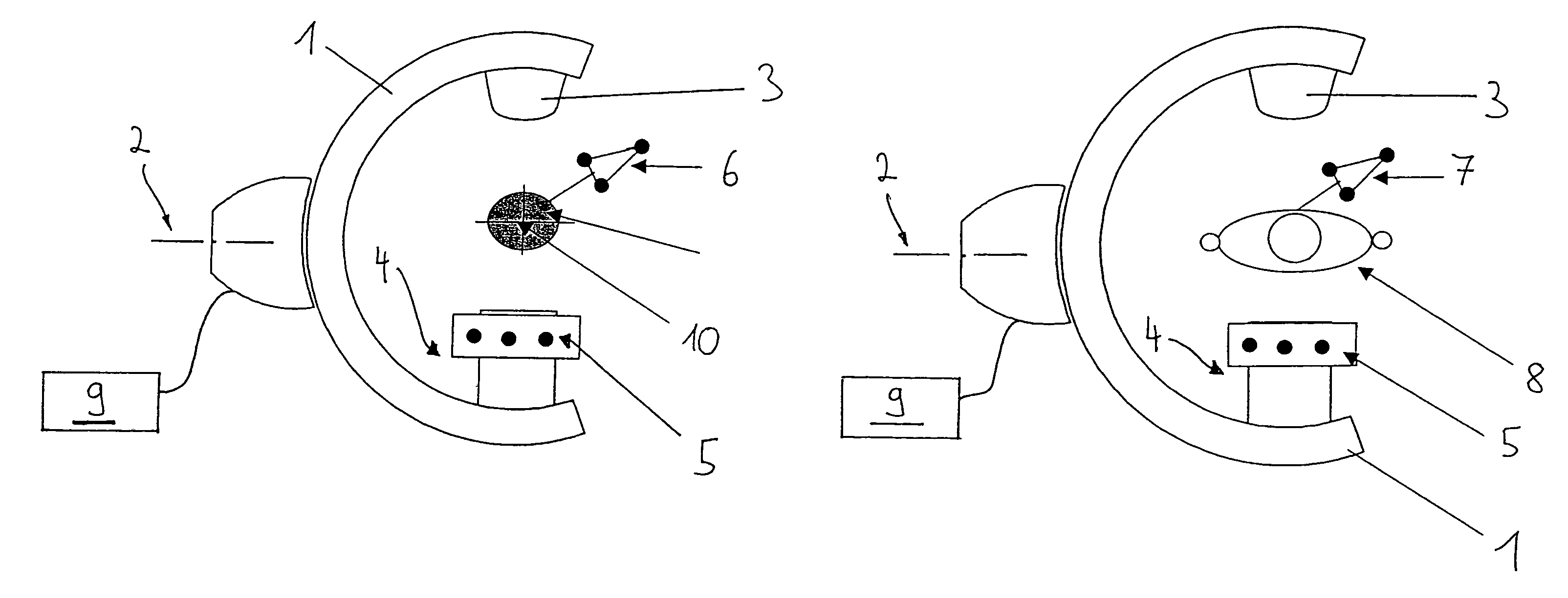
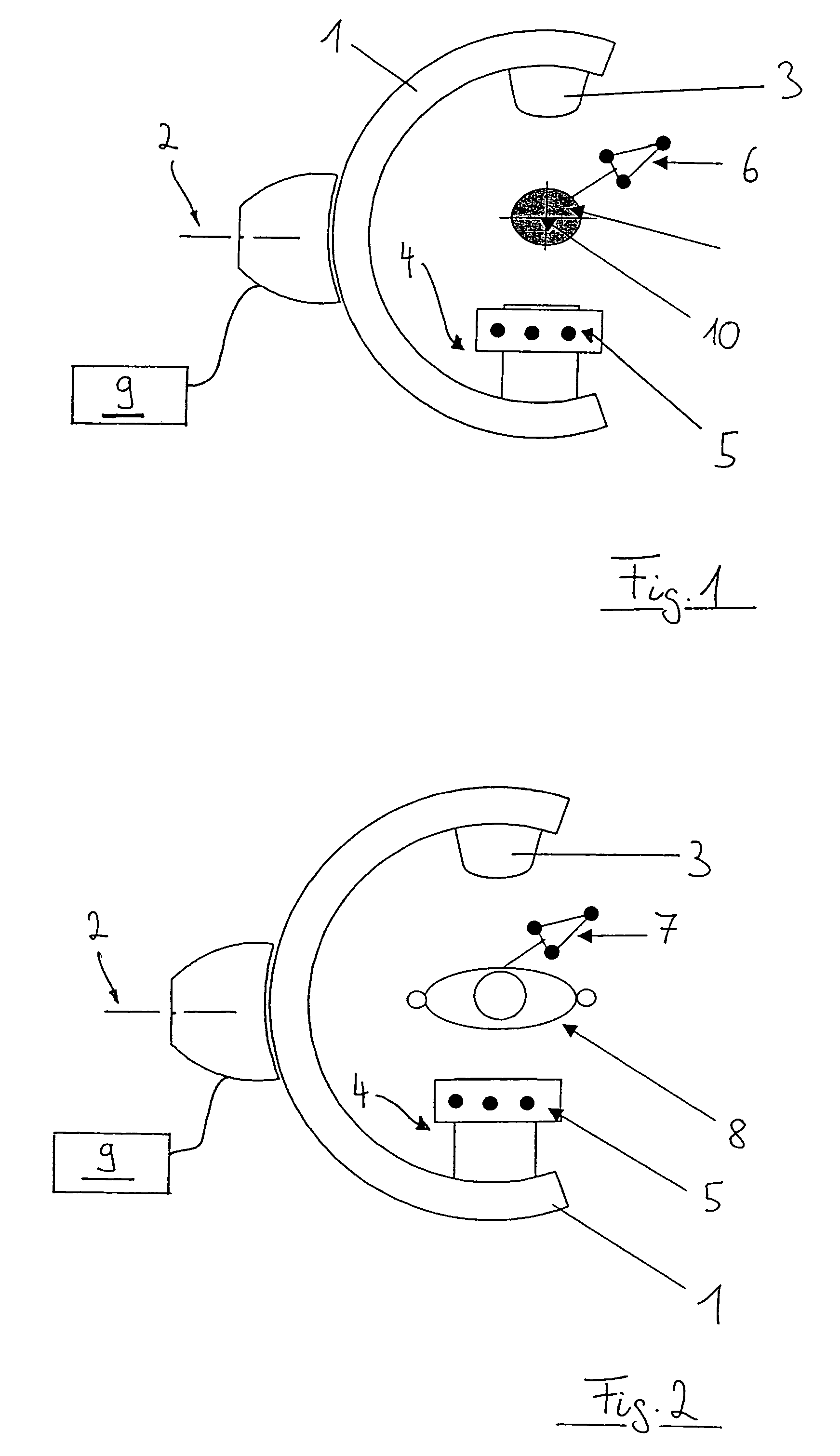
[0020]With reference to FIG. 1, a C-arm x-ray device 1 includes an arm, which can rotate about an axis 2 for recording x-ray images, such that, for example, a series of isocentric x-ray image recordings can be produced. The C-arm x-ray device 1 includes a radiation source 3 on an upper side and an image recorder 4 on an opposite side. In one embodiment, the image recorder 4 supports a calibration attachment, which can include a first marker geometry 5. A navigation system 9, which is shown schematically, can positionally detect and track positions of the C-arm (for example, via the marker geometry 5), and positions of objects present in the x-ray device radiation field. For example, the navigation system 9 can detect and track positions of a calibration phantom 10 (via its associated marker geometry 6), as shown in FIG. 1, or a patient 8 (via its associated marker geometry 7), as shown in FIG. 2. This position detection can be accomplished, for example, using cameras. Alternatively,...


PUM


Abstract

A method for displaying images in a medical navigation system assisted by x-ray images includes calibrating an x-ray device in the medical navigation system and producing a plurality of two-dimensional x-ray images of a patient using the x-ray device. During the producing step, positions of the x-ray device are determined using the medical navigation system in order to produce positional data. Data associated with the two-dimensional x-ray images is converted into three-dimensional data. The two-dimensional x-ray images, production information corresponding to the two-dimension x-ray images, and the three-dimensional data to the navigation system. At least the two-dimensional x-ray images are displayed on an image output of the medical navigation system.

Description

RELATED APPLICATION DATA[0001]This application claims priority of U.S. Provisional Application No. 60 / 440,702, filed on Jan. 17, 2003, which is incorporated herein by reference in its entirety.FIELD OF THE INVENTION[0002]The present invention relates to x-ray image-assisted navigation in the area of medicine. It provides a method for displaying images in a medical navigation system assisted by x-ray images.BACKGROUND OF THE INVENTION[0003]Modern, computer-assisted surgery is able, with the aid of medical navigation systems, to show the surgeon's instruments or implants in relation to anatomical image data on a monitor during the operation. The anatomical image data of a patient can be acquired either pre-operatively or intra-operatively and can consist of various modalities (CT, MRI, PET, SPECT, x-ray images or combinations of these).[0004]In computer-assisted surgery, it is also common to ascertain image data with the aid of an x-ray device, generally used in the form of a C-arm, a...

Claims


Application Information

Patent Timeline
IPC IPC(8): A61B5/05A61B19/00
CPCA61B19/5244A61B2019/5238A61B2019/5255A61B2019/5272A61B34/20A61B2034/2055A61B2034/2072A61B2090/376
Inventor ESSENREITER, ROBERTSEIFFERTH, FALKOSCHMIDT, ROBERTZEISS, MARIONEAGU, SORIN-ALEXANDRUMUHLHAUSSER, MATTHIASSTEINER, ANDERS
Owner BRAINLAB
Why Eureka
Try Eureka