Intramedullary implant for fracture fixation

a technology of intramedullary implants and fractures, which is applied in the field of intramedullary implants for fracture fixation, can solve the problems of high deformation load across the fractured articular surface, loss of reduction of joint surface, and malposition of wrist, and achieves the effects of reducing the risk of failure, and maintaining the length of fragments

Inactive Publication Date: 2007-09-11
MEDOFF ROBERT J +2
View PDF4 Cites 56 Cited by
  • Summary
  • Abstract
  • Description
  • Claims
  • Application Information

AI Technical Summary

Benefits of technology

[0014]An object of the present invention is to provide an implant adapted to overcome the limitations noted above by placing a large surface area of contact at the tip of a buttressing element against an apical corner of a subarticular fragment, such as the radial styloid. The tip of the buttressing element of the implant is configured with a blunt shape to supports the surface geometry of the subchondral bone of a fracture element from inside the osseous structure of the bone fragment. This loads the implant axially, and eliminates torque between the bone and implant, reducing the risk of failure. In addition, since the bone is suspended from the tip of the implant, like a tent being held by a tent post, fragment length is maintained.
[0015]In one embodiment, the end of the buttressing element is in the form of a smooth rounded tip of bullet-like shape in order to fit inside the radial styloid and prevent loss of length. The tip of the buttressing element is impacted against the endosteal side of the apical contour of the end of the radial styloid in order to hold it out to length; proximally, the buttressing element exits the bone and is fixed with screws to the radial shaft to secure it into position. The shape of the tip may be symmetrical or asymmetrical depending on the configuration of the apical contour.
[0016]Additional features of the invention provide improved insertion technique as well as the quality of fixation. One embodiment simplifies insertion by adding a cannulated track through the length of the implant to allow the implant to be placed over a guide wire. In another embodiment straight or angled crossing holes are formed in the implant in order to allow screws, posts or pins to be placed as locking fasteners across the tip of the implant from outside the bone, penetrating the implant like the locking screws on an interlocking nail. In further modifications of this latter embodiment, the crossing holes are threaded so that a crossing post can be inserted from outside the bone using an insertion guide that is attached to the proximal end of the implant. The crossing post is threaded and has a head secured on a washer. As it is locked across the tip of the implant within the radial styloid, an additional buttress radially is provided, preventing displacement of the radial column radially and preventing widening of the articular surface.
[0020]The buttressing element can be coupled to a dorsal or volar plate, using an angled base that redirects the connection to the volar or dorsal side, yet still providing an implant that achieves a buttress effect at the tip of the buttressing element.
[0021]According to one aspect of the invention, the implant serves for fixation of a fracture of the radius and comprises an elongated implant having a proximal portion adapted for connection to stable bone and a distal portion adapted for buttressing the radial styloid of an unstable bone fragment of the fracture. The distal portion is shaped for insertion into an intramedullary canal of the unstable bone fragment to enter the radial styloid and provide a broad buttress support for a tip end of the radial styloid at the endosteal surface thereof.
[0024]According to this aspect of the invention, the implant comprises a first part for fixation to a surface of a first bone fragment, and a second part for insertion within a second bone fragment, the second part having a tip end adapted to provide a broad abutment surface against an endosteal surface at a conical end of the second fragment to maintain length of the second bone fragment relative to the first for preventing axial collapse or shortening of the second bone fragment. The second or buttressing part can be located distally or proximally depending on the location of the fragment with the conical end.

Problems solved by technology

If the radial styloid is not held out to length, the bones of the carpus (wrist) will subside proximally and result in high deforming loads across the fractured articular surface.
This can lead to loss of reduction of the joint surface and malposition of the wrist.
In addition, since the adjacent ulna is of normal length and the radius becomes short, significant dysfunction can occur to the adjacent radioulnar joint.
Finally, translation of the radial styloid to the radial side causes the end of the articular surface to spread apart, resulting in incongruity of the articular surface and a poor clinical result.
Current implants do not adequately address this problem of maintaining length to the radial styloid.
In addition, since the pins that cross the radial styloid are thin, the fragment can slide along the pins and lose length.
Standard buttress fixation plates placed either dorsally or volarly also do not adequately address this problem.
These also require exposure of the entire surface of the bone, which can be detrimental because of stripping of the blood supply and irritation of the soft tissues.
This type of approach requires a bulky plate to be placed on the superficial surface of the distal fragment in a region where many tendons and ligaments are in close apposition to the bone; this can result in tendon irritation and even rupture.
In addition, because these plates are placed on the dorsal or volar surface, the only means available for supporting the distal articular surface is limited to cylindrical posts or screws that are placed through holes in the plate.
Since these posts or screws are designed to cross the bone either from dorsal to volar or from volar to dorsal, it is not geometrically possible to angle the tip of a post or screw into the apical corner of the radial styloid using a dorsal or volar plate.
If this occurs, the screw or post is placed under torque, increasing the forces in both the implant and bone / implant interface.
This torque may lead to loosening and make the bone fragment more prone to slip off the fixation post.
Increased stresses in the bone implant interface also increase the risk of failure.
Finally, because of the complex geometry and intimate apposition of tendons and ligaments against the volar and dorsal surfaces of the radius, trying to secure a distal radial styloid fragment using dorsal or volar fixation is prone to complications and is generally not used.
However, these implants are intentionally designed to allow sliding and impaction of the femoral head to promote union; in this circumstance the loss of length is a minor problem compared to the possibility of non-union by maintaining length.
In addition, with the existing hip screws and blade plates, no mechanism is present to buttress the subchondral bone of the femoral head with a smooth tip.

Method used


Image

  • Intramedullary implant for fracture fixation
  • Intramedullary implant for fracture fixation
  • Intramedullary implant for fracture fixation

Examples


Embodiment Construction

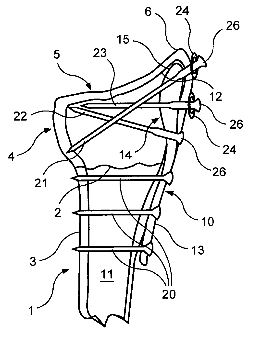
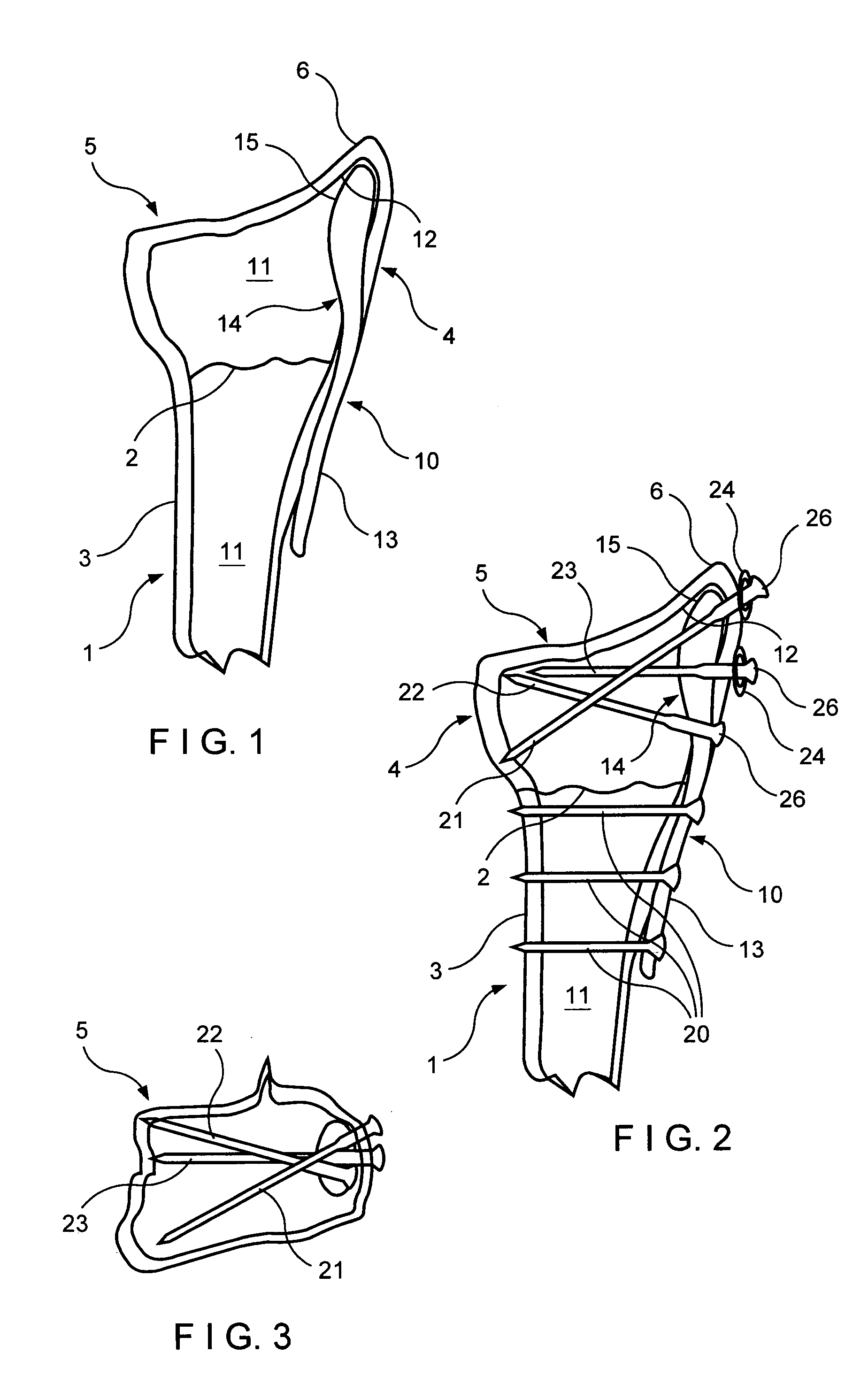
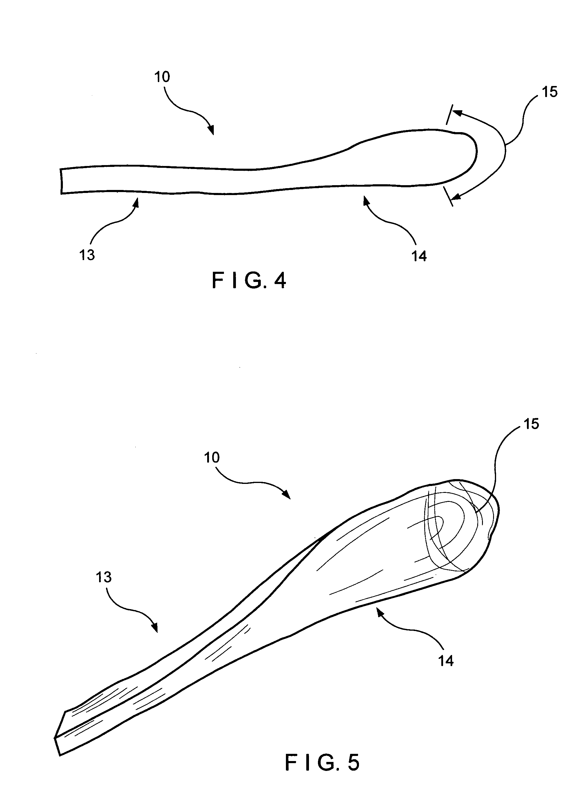
[0037]The invention will be described hereafter with reference to fixation of a fracture of the radius of the wrist utilizing an intramedullary implant which is braced against the apex of the lower tip of the radial styloid to serve as a prop or buttressing means to hold an unstable fragment of the fracture at length to prevent shortening. However, as will be evident to those skilled in the art, the invention is applicable to other fractures as well, in which the implant can be inserted into an intramedullary canal and braced in an inner apical surface of a bone fragment adjoining the fracture. Suitable examples are the distal end of the fibula, the medial malleolus of the ankle and the distal end of the ulna.

[0038]Referring to FIG. 1, therein is seen a bone 1, namely the radius of the wrist, in which a fracture 2 is present at its distal end and forms a stable proximal bone fragment 3 and an unstable distal bone fragment 4. The distal bone fragment 4 has a distal end 5 which includ...


PUM


Abstract

An implant for fracture fixation having external portion shaped to be externally engaged on and secured to a stable bone fragment on one side of a bone fracture and an integral internal portion sized and shaped for being inserted into and accommodated in an intramedullary canal of an unstable bone fragment on an opposite side of the fracture. The internal portion has an end with a rounded tip of bullet-shape for fitting in an apical space at an endosteal surface of the unstable bone fragment to provide a bearing support for the unstable bone fragment over a large area.

Description

FIELD OF THE INVENTION[0001]The invention relates to fracture fixation devices and particularly to devices for fixation of distal fractures of the radius.[0002]In particular, the invention relates to an intramedullary implant for such fracture fixation in which the implant includes a first portion adapted to be secured externally on stable bone and a second portion which enters into the intramedullary canal of the unstable bone fragment to be set and wherein the tip end of the second portion is shaped to fit in a space in an endosteal surface of the unstable bone fragment and suspend the unstable bone fragment by said tip end to prevent axial collapse or shortening of the unstable bone fragment.[0003]In the case of the fracture of the distal radius, the invention provides that the tip end of the intramedullary implant engage the subchondral bone of the radial styloid to maintain its length and position.[0004]The invention further relates to a method for fracture fixation with an int...

Claims


Application Information

Patent Timeline
Patent Type & Authority Patents(United States)
IPC IPC(8): A61B17/58A61B17/17A61B17/72A61B17/80A61B17/86
CPCA61B17/1721A61B17/1725A61B17/1728A61B17/72A61B17/8061A61B17/8695A61B2017/1782A61B17/1782
Inventor MEDOFF, ROBERT J.
Owner MEDOFF ROBERT J
Why Eureka
Try Eureka