Medical injection system

Active Publication Date: 2007-12-11
ACIST MEDICAL SYST
View PDF60 Cites 90 Cited by
  • Summary
  • Abstract
  • Description
  • Claims
  • Application Information

AI Technical Summary

Benefits of technology

[0036]Because the automatic injector is driven by at least one microprocessor, the system must be capable of storing the data and software used for executing the application. It would be desirable to have the capability to install software after the device has been assembled. This capability facilitates ease of manufacture and allows immediate field upgrades without significant down time. Thus, it is preferable to provide the software and data storage capability on a modular memory card, such as CompactFlash™. The CompactFlash™ mass storage device is a card which can be unplugged and replaced through an access point on the injector devi

Problems solved by technology

Thus, if extravasation occurs while injecting

Method used


Image

  • Medical injection system
  • Medical injection system
  • Medical injection system

Examples


Embodiment Construction

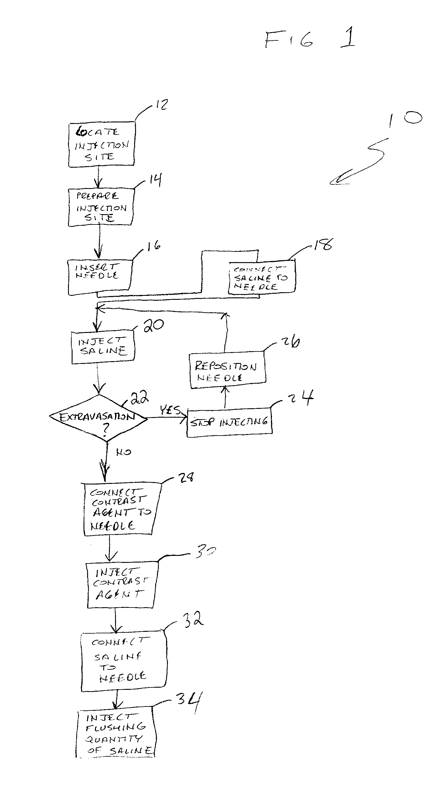
[0057]Method of Preventing Extravasation

[0058]FIG. 1 shows a flow diagram of the method of preventing extravasation 10 of the present invention. Beginning at 12, an injection site is located by the attending health professional and prepared for injection at 14 using appropriate cleaning techniques. The needle or catheter is inserted at 16 to establish fluid communication between the needle or catheter and the targeted lumen of the patient.

[0059]At 18, a supply of saline is fluidly connected to the needle or catheter and, at 20, a quantity of saline is injected into the patient at a predetermined flow rate that may be approximately equal to the desired flow rate of the eventual contrast agent injection. It is preferred that the flow rate of the saline injection be at least as great as the planned flow rate of the contrast agent. Doing so ensures that extravasation complications caused by jetting forces will be revealed prior to the introduction of the contrast agent. While the saline...


PUM


Abstract

A method of preventing extravasation of contrast agent during a computed tomography injection. An automatic injector device facilitates ease of accomplishing the method. The method includes establishing the absence of extravasation using an absorbable injectate, such as saline, prior to injecting the contrast agent. The device includes a computerized injector head capable of switching between two injectates without physical human intervention. The device is controlled by a remote operating panel located in a control room that is protected from X-ray radiation. The device includes various software driven safety features that prevent the occurrence of unsafe conditions.

Description

CROSS REFERENCE TO RELATED APPLICATIONS[0001]This application claims priority from U.S. Provisional Application Serial No. 60 / 294,471 filed on May. 30, 2001 and entitled CT INJECTOR SYSTEM, incorporated by reference herein in its entirety.BACKGROUND OF THE INVENTION[0002]Computed tomography (hereinafter “CT”) is a medical procedure whereby an X-ray imaging machine is used to take cross-sectional images of a patient. The source of the X-rays is placed on one side of the body while an array of detectors is placed on the other side. The X-rays pass through the body and are read by the detectors on the other side. The signals received by the detectors are sent to a computer which compiles the data to create images. The detectors and X-ray source may be rotated around the body, while the body is being translated axially, to create a plurality of layered images.[0003]CT differs from traditional X-ray imaging in that a computer is used to first “record” the image. Often, a contrast agent p...

Claims


Application Information

Patent Timeline
IPC IPC(8): A61M5/00A61M5/145A61M5/19A61M5/31A61M5/315
CPCA61M5/007A61M5/14546A61M5/14566A61M2005/3128A61M5/31513A61M5/31515A61M5/19
Inventor TOEWS, GLENNSZCZECH, CHRISMCPEAK, THOMAS J.DUCHON, DOUGLAS J.
Owner ACIST MEDICAL SYST
Why Eureka
Try Eureka