
Method of full-color optical coherence tomography

a tomography and color technology, applied in the field of biomedical imaging, can solve the problems of mechanical scanning being a major limitation factor in placing a fundamental limit on the image acquisition speed, and compromising the lateral resolution to a few m, and achieve the effect of high frame ra

Inactive Publication Date: 2008-01-08
UNIV OF SOUTH FLORIDA
View PDF2 Cites 27 Cited by
  • Summary
  • Abstract
  • Description
  • Claims
  • Application Information

AI Technical Summary

Benefits of technology

[0013]In accordance with the present invention a novel method of optical tomography for surface and sub-surface imaging of biomedical tissues is provided. The method is based on the principle of wide field optical coherence tomography (OCT) and is capable of providing full-color three-dimensional views of a tissue structure with high-resolution and high frame rate.
[0015]In an additional embodiment, a novel method of full color two-dimensional optical coherence tomography is provided for surface and sub-surface imaging of biomedical tissues. The method as described is based on the principle of wide field optical coherence tomography and is capable of providing full-color two-dimensional and three-dimensional images of a tissue structure with high-resolution and high frame rate.

Problems solved by technology

The three-dimensional image is constructed by pixel-by-pixel mechanical scanning of the entire object volume, which places a fundamental limit on the image acquisition speed.
In order to maintain the high speed of the z-scan, a considerably large depth of field, approximately a mm, is needed, which compromises the lateral resolution to a few μm.
On the other hand, the mechanical scanning is a major limiting factor in the image acquisition speed.
In CSM, such 2D imaging is approximated with a large number of well-spaced apertures, such as rotating Nipkow disk or multi-aperture scanning using an electro-optic spatial light modulator, but the light efficiency or image contrast tends to be low.

Method used


Image

  • Method of full-color optical coherence tomography
  • Method of full-color optical coherence tomography
  • Method of full-color optical coherence tomography

Examples


Embodiment Construction

[0031]In the following detailed description of the preferred embodiments, reference is made to the accompanying drawings, which form a part hereof, and within which are shown by way of illustration specific embodiments by which the invention may be practiced. It is to be understood that other embodiments may be utilized and structural changes may be made without departing from the scope of the invention.

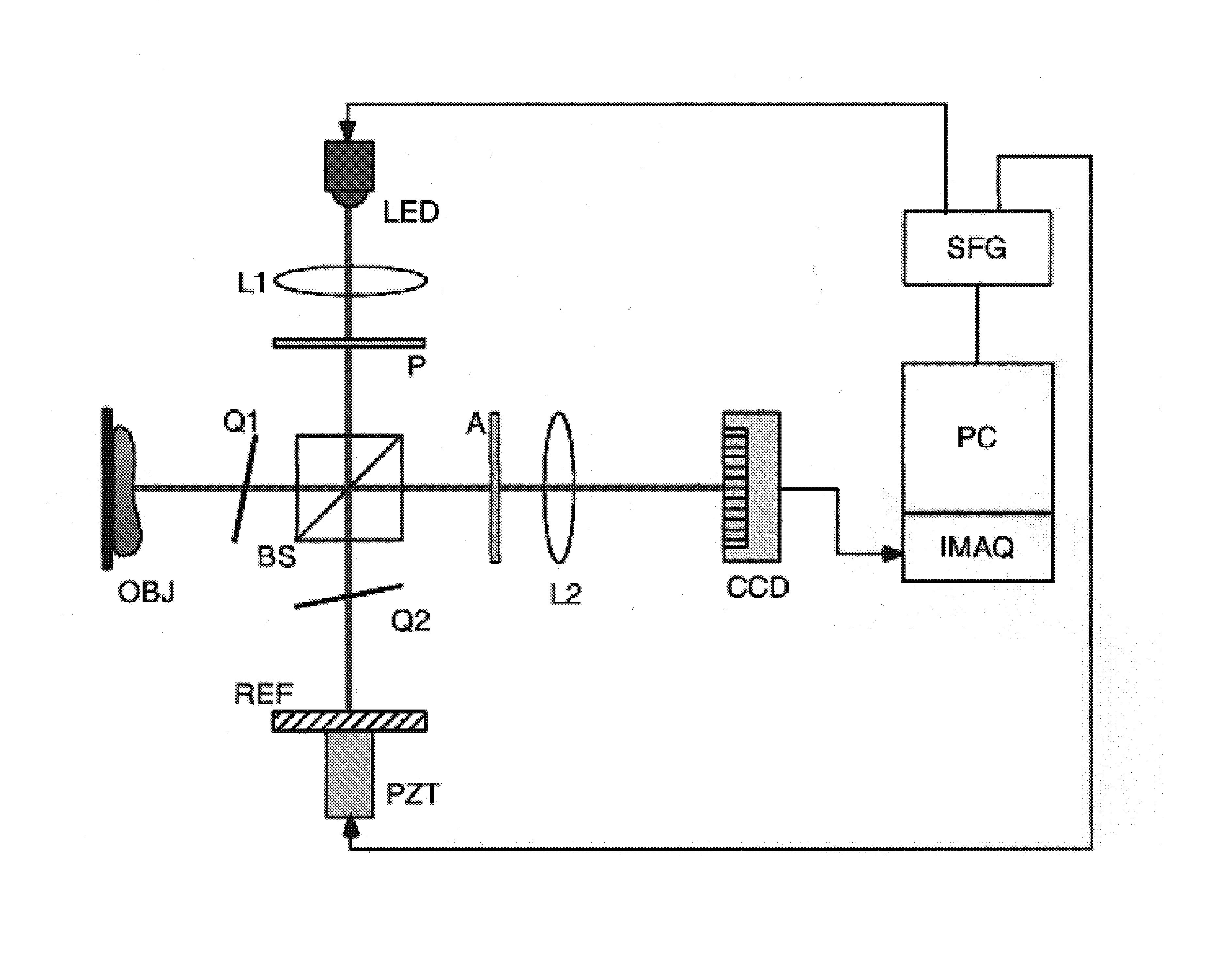
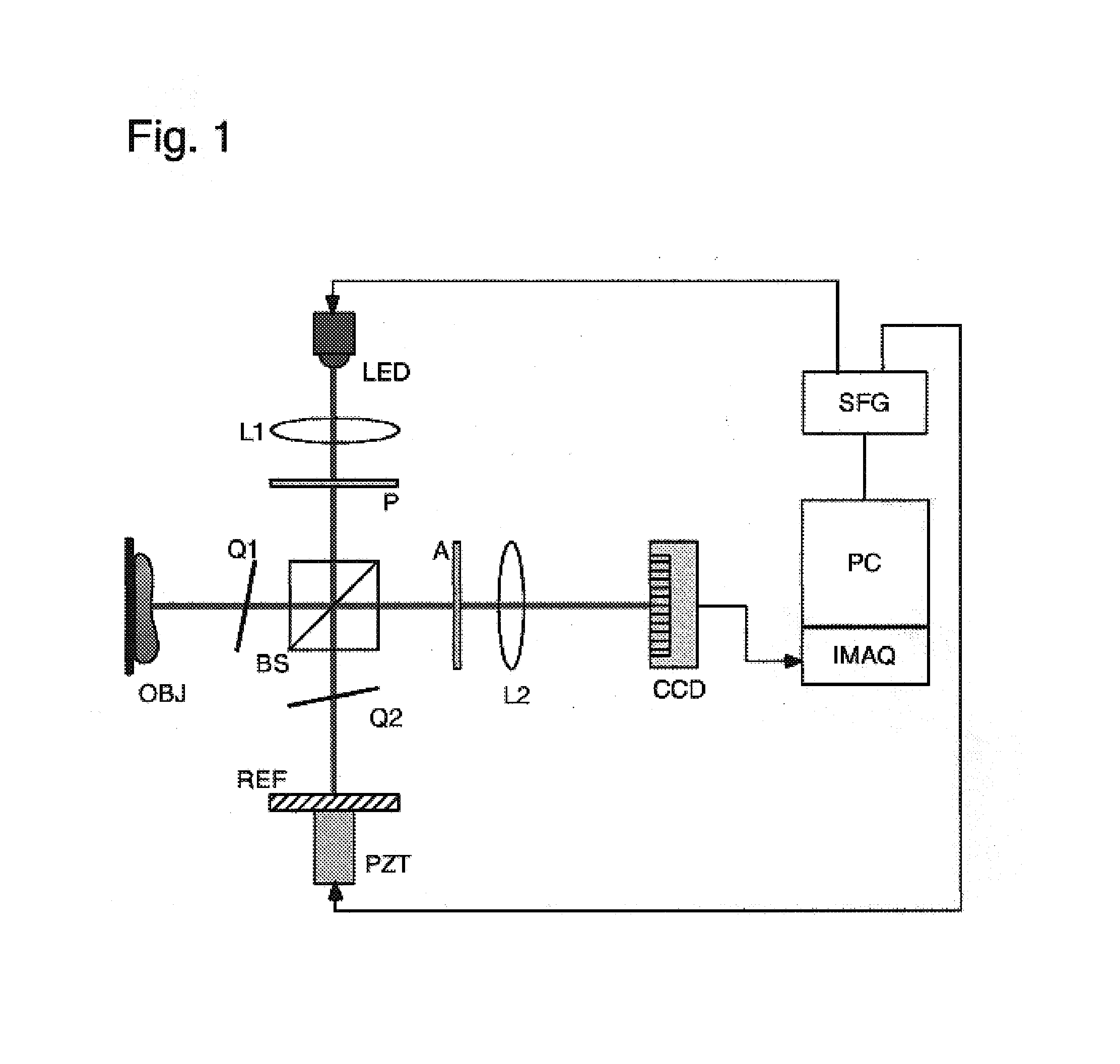
[0032]The principle of color 3D microscopy by wide-field optical coherence tomography (WFOCT) is described referring to the diagram of the apparatus illustrated in FIG. 1. A high-brightness LED (˜30 lumens) illuminates the Michelson interferometer through a collimating lens L1, a polarizer P, and the broadband polarizing beam splitter BS. The quarterwave plates Q1 and Q2 in the object or reference arms change the orthogonal polarization states, so that all of the reflections from the object or the reference mirror are steered toward the monochrome CCD camera through the imaging lens ...


PUM


Abstract

Two-dimensional and three-dimensional optical coherence tomography is obtained by differential imaging of full-frame interference images using a white light source. Full-color tomographic imaging is also possible by processing the three-color channels of the interference images. A technique is described to obtain two-dimensional OCT images with full natural color representation. In a particular embodiment, the interference image is acquired using a color camera and the three-color channels are processed separately, recomposing the final image. In an additional embodiment, the interference images are acquired using separate red, blue and green light sources and the three color channels are combined to recompose the final image.

Description

CROSS-REFERENCE TO RELATED APPLICATIONS[0001]This application is a continuation of prior application Ser. No. 11 / 063,499 filed on Feb. 22, 2005, now U.S. Pat. No. 7,095,503 B2, the contents of which are herein incorporated by reference, which application claims the benefit of priority to provisional Application No. 60 / 546,408 filed Feb. 20, 2004, entitled “Method and Apparatus for Full-Color Two-Dimensional Optical Coherence Tomography,” and to provisional Application No. 60 / 635,162 filed Dec. 10, 2004 entitled “Full-Color Three-Dimensional Microscopy by Wide-Field Optical Coherence Tomography.”STATEMENT OF GOVERNMENT INTEREST[0002]This invention was made with Government support under Grant No. DBI 0243237 awarded by the National Science Foundation. The Government has certain rights in the invention.BACKGROUND OF INVENTION[0003]In the field of biomedical imaging, various methods of optical imaging of biomedical tissues known in the art offer significant advantages over other biomedi...

Claims


Application Information

Patent Timeline
IPC IPC(8): G01B11/02
CPCG01N21/4795G01B9/02007G01B9/02091G01B9/02087G01N21/49G01N2021/1787G01B2290/70
Inventor KIM, MYUNG K.YU, LINGFENG
Owner UNIV OF SOUTH FLORIDA
Learn More