Simultaneous multi-headed imager geometry calibration method

a multi-headed imager and calibration method technology, applied in the field of spect imaging of small animals, can solve the problems of loss of this important dynamic information, errors (artifacts) showing up in the reconstructed image, and the technique requires complicated system modeling and assumptions about the dynamic, and the special clinical spect system is at present limited to imaging the brain

Inactive Publication Date: 2008-02-19
JEFFERSON SCI ASSOCS LLC
View PDF3 Cites 27 Cited by
  • Summary
  • Abstract
  • Description
  • Claims
  • Application Information

AI Technical Summary

Problems solved by technology

However, most of the body functions are dynamic and this leads to the loss of this important dynamic information and even to errors (artifacts) showing up in the reconstructed images.
However, these techniques require complicated system modeling and assumptions about the dynamic processes that are to be unfolded in 3d reconstructions from the limited dynamic angular data obtained in a normal clinical acquisition with a slowly rotating gamma cameras.
However, these special clinical SPECT systems are at present only limited to imaging the brain.
This leads to problems with image reconstruction, such as artifacts, as mentioned above.
In this way, the useful field-of-view is highly reduced and limited (by design) to primarily accurately image animal organs, such as the brain, heart, etc, and only a whole small mouse can be imaged.
Therefore, that system cannot provide a dynamic image of a larger whole rodent animal such as a rat.
In addition to the fixed size issue, the stationary SPECT design of Barrett et al uses few pinholes (one per module) and therefore cannot be optimized for high efficiency.
In our parallel-hole close geometry configuration it would not be possible to place even that small a number of modules on a ring and close to an animal.
This complicated concept cannot be easily adapted to a larger FOV such as a whole rat, or even a mouse.
While offering good dynamic performance for the above clinical applications on human subjects, the system does not exhibit high enough spatial resolution to be used on small animals.
As a result, spatial resolution on the central axis is only 3 mm FWHM, which is only marginally applicable to small animal imaging.
The tilting, swiveling and translating motions, are to collect more tomographic projections to assure higher quality imaging and it has merit in the clinical situations with sick bed-ridden patients etc, but this is a rather complicated and not well optimized approach for small animal imaging application.
Standard reconstruction techniques do not allow temporal information to be obtained from this inconsistent pr

Method used


Image

  • Simultaneous multi-headed imager geometry calibration method
  • Simultaneous multi-headed imager geometry calibration method
  • Simultaneous multi-headed imager geometry calibration method

Examples


Embodiment Construction

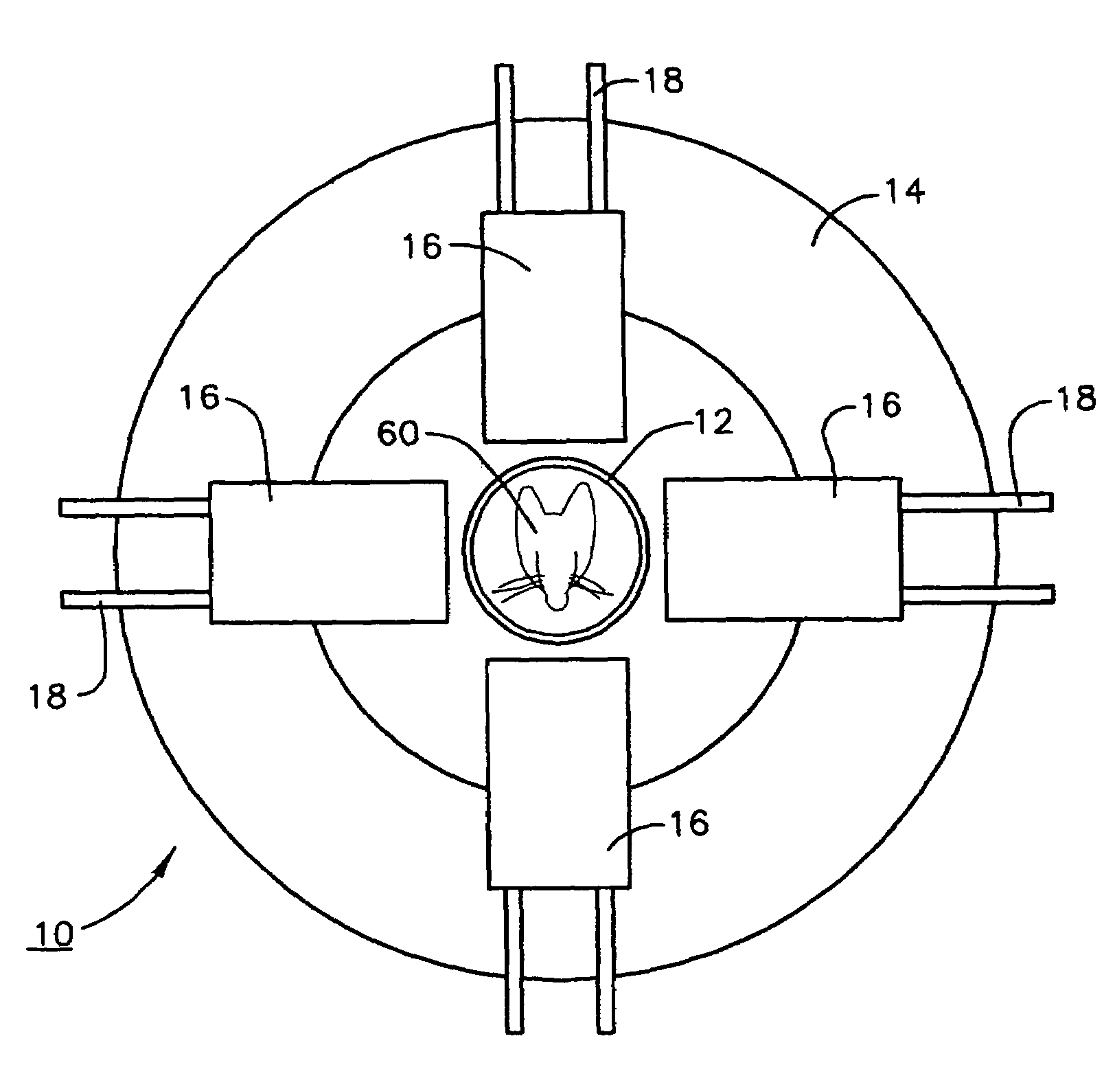
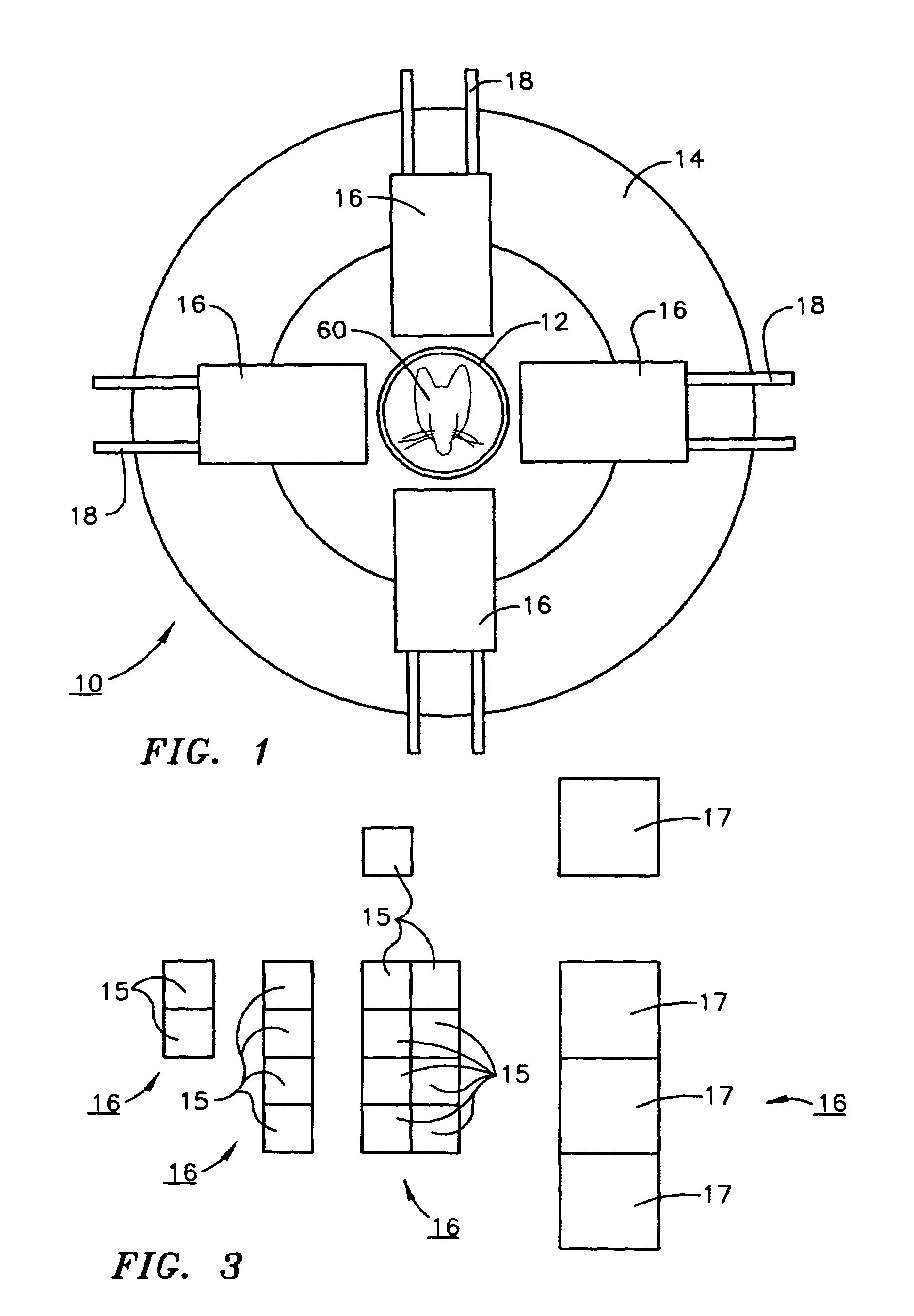
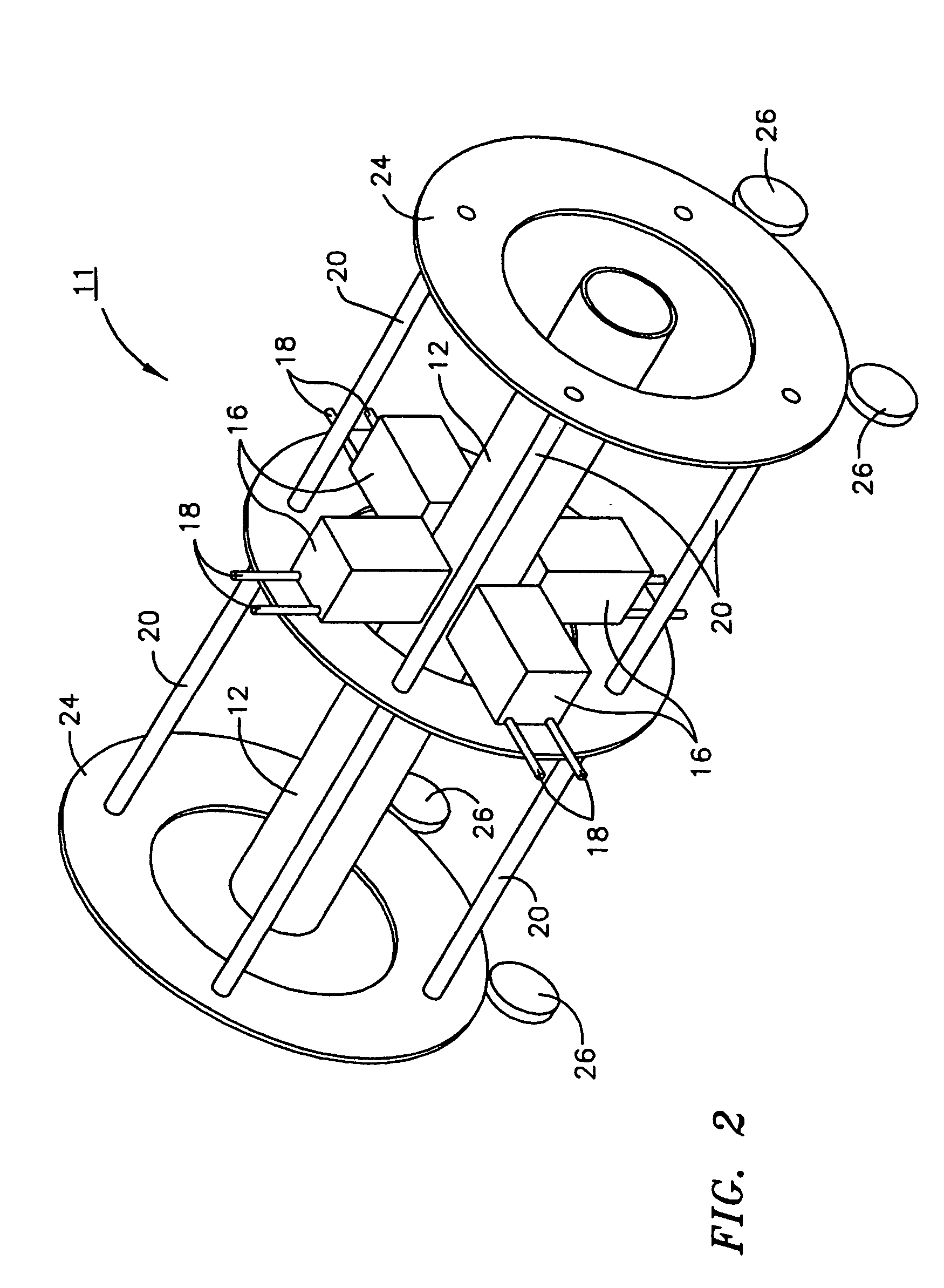
[0044]What follows, is a detailed description of one SPECT multi-headed imager system in which the calibration system described herein will find use. While the calibration system of the present invention is described primarily in connection with such a multi-headed SPECT imaging system that finds use in the study of small animals, it should be noted that the calibration system of the present invention will be useful in the calibration of any multi-headed imaging system, PET, X-ray, etc. that rotates about a subject under study and utilizes imaging heads that are “gantry” mounted.

[0045]A knowledge of the dynamic biodistribution on a short time scale of minutes or even seconds of an injected or otherwise animal body administered imaging biomarker used either in diagnostics or in treatment, is crucial in many biological studies requiring measurement of blood flow distribution after biomarker injection, and subsequent uptake and washout by all organs, for example, in studies of the toxi...


PUM


Abstract

A method for calibrating multi-headed high sensitivity and high spatial resolution dynamic imaging systems, especially those useful in the acquisition of tomographic images of small animals. The method of the present invention comprises: simultaneously calibrating two or more detectors to the same coordinate system; and functionally correcting for unwanted detector movement due to gantry flexing.

Description

[0001]The United States of America may have certain rights to this invention under Management and Operating Contract No. DE-AC05-84ER 40150 from the Department of Energy.FIELD OF THE INVENTION[0002]The present invention relates to the SPECT imaging of small animals and more particularly to calibration methods useful for obtaining high resolution, high efficiency dynamic images of small animals injected with a biomarker.BACKGROUND OF THE INVENTION[0003]Single Photon Emission Computed Tomography (SPECT) as a form of nuclear medicine imaging is used to show how organs, systems and different molecular processes in the human body are functioning. Acquisition of 3d SPECT images takes about 20-40 minutes, depending on procedure, and during this time it is usually assumed that the ‘function’ that is being imaged is stationary, not changing in time during the duration of the scan. During the SPECT scan the 1 to 3 imaging detector heads are rotating slowly around the patient recording views o...

Claims


Application Information

Patent Timeline
IPC IPC(8): G01T1/161
CPCA61B6/037A61B6/501A61B6/508G01T1/1644
Inventor TRAN, VI-HOAMEIKLE, STEVEN RICHARDSMITH, MARK FREDERICK
Owner JEFFERSON SCI ASSOCS LLC
Why Eureka
Try Eureka