Breast cancer detection system

a breast cancer and detection system technology, applied in the field of medical diagnostic imaging, can solve the problems of reducing the accuracy of conventional breast cancer screening methods, affecting so as to avoid unnecessary generic models, improve the accuracy and overcome the deficiency of conventional confocal microwave imaging.

Active Publication Date: 2008-03-04
UNIV OF FLORIDA RES FOUNDATION INC
View PDF10 Cites 32 Cited by
  • Summary
  • Abstract
  • Description
  • Claims
  • Application Information

AI Technical Summary

Benefits of technology

[0013]The present invention relates to the field of microwave imaging system for breast cancer detection. Using one embodiment of the present invention, the deficiencies of conventional confocal microwave imaging can be overcome in that neither a cylindrical nor a planar modeling configuration for breasts is necessary. Rather, in accordance with the inventive arrangements, the microwave imaging system can produce accurate detection of small to large tumors in the breast with specific reference to the shape, size, and boundary irregularities of the breast under study. Importantly, so as to avoid the inaccuracies of the planar and cylindrical configurations, variably positioned probing antennae can be affixed to the breast surface. The actual position of the probing antennae can be determined through the use of a fixed position array of passive antennae. In this way, the breast can be modeled in accordance with the actual size and shape of the breast under study, and unnecessary generic models can be avoided, thereby increasing the accuracy of tumor detection over conventional methodologies.

Problems solved by technology

Though advances in the treatment of breast cancer have been made, the effectiveness of conventional breast cancer screening methods remains questionable.
Specifically, a decade-long study conducted amongst 260,000 Chinese women in Shanghai concluded that, in general, women examining their breasts remain unable to detect tumors early enough to reduce their risk of dying from breast cancer.
In particular, the effectiveness of x-ray mammography has been questioned at all levels.
In this regard, much of problem with x-ray mammography relates to the inability for x-ray mammography to image dense tissue.
Consequently, the results of an x-ray mammography may show suspicious areas where no malignancy exists (up to twenty percent of biopsied growths identified as cancerous by a mammogram are identified as malignancies).
Finally, the current process for conducting x-ray mammography can be severely uncomfortable.
Specifically, the use of imaging plates can cause bruising in the breasts and can, therefore, be a significant disincentive for women to undergo mammography screening.
Nevertheless, confocal microwave imaging techniques for breast cancer detection remain unable to precisely differentiate malignancies from benign tumors, largely due to the imprecise modeling of the breast.
Moreover, irregularities can persist about the boundary between the breast skin and surrounding tissue and also about the boundary between the breast tissue and the chest wall.

Method used


Image

  • Breast cancer detection system
  • Breast cancer detection system
  • Breast cancer detection system

Examples


Embodiment Construction

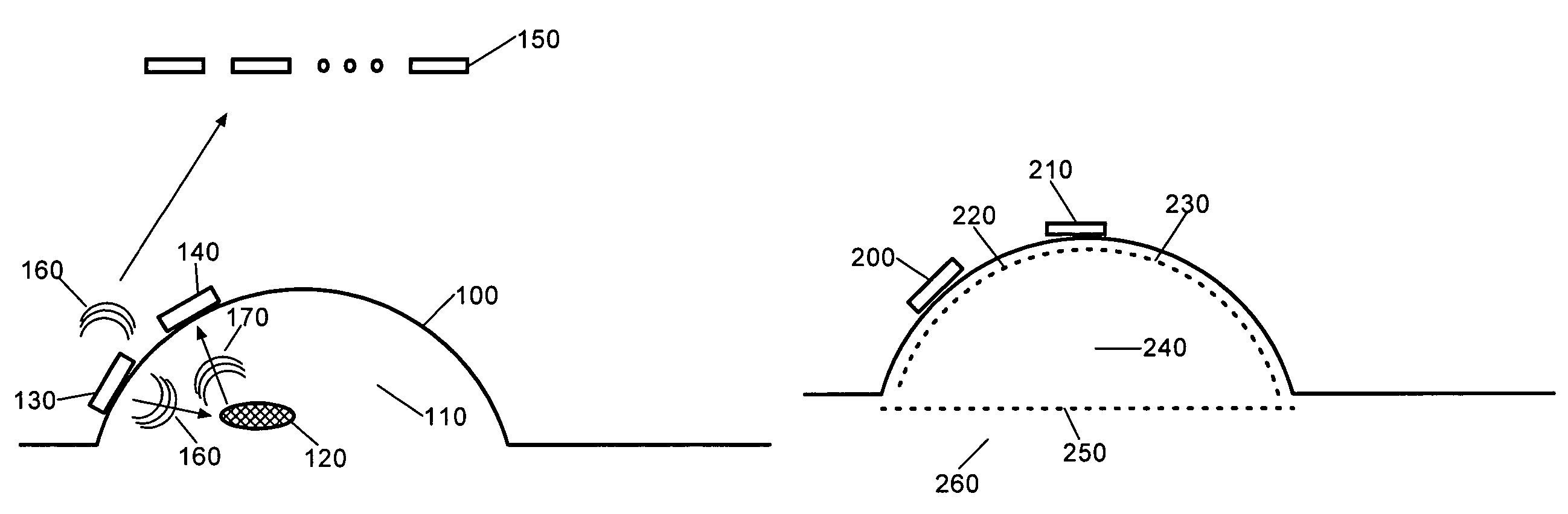
[0023]The present invention provides a microwave imaging process and system for use in breast cancer detection. The process can include the steps of transmitting microwave energy from a first probing antenna at the surface of the breast inwardly through the breast tissue, receiving at a second probing antenna at the surface of the breast reflected ones of the transmitted microwave energy which had been reflected by a tumor disposed in the breast tissue, and further receiving in an array of fixed position antennae emissions from said transmitted microwave energy. The position of the probing antennae can be computed based upon position location techniques applied to the time of receipt of the emissions. Based upon computed position, an image of the tumor in the breast tissue can be formed.

[0024]Importantly, clutter reduction can be applied to the formation of the image of the tumor so as to reduce the effect of microwave energy reflected not by the tumor, but by the boundaries between...


PUM


Abstract

A method for detecting breast cancer can include the step of positioning a transmitting antenna and a receiving antenna about a breast so that the transmitting antenna is positioned to transmit microwave energy into the breast and the receiving antenna is positioned to receive the transmitted energy after the energy has passed into the breast. Microwave energy can be transmitted from the transmitting antenna. The microwave energy can be received by the receiving antenna and used to determine a presence of tumors within the breast. A relative position of the transmitting antenna and the receiving antenna can be adjusted about the breast. After each position adjustment, the transmitting and the receiving steps can be repeated.

Description

CROSS REFERENCE TO RELATED APPLICATIONS[0001]This application claims the benefit of U.S. Provisional Application No. 60 / 447,487, filed in the United States Patent and Trademark Office on Feb. 14, 2003, the entirety of which is incorporated herein by reference.BACKGROUND[0002]1. Statement of the Technical Field[0003]The present invention relates to the field of medical diagnostic imagery and, more particularly, to breast cancer detection systems.[0004]2. Description of the Related Art[0005]Breast cancer is the most common major cancer among women in the United States and breast cancer is the leading cause of cancer deaths in women, second only to lung cancer. An estimated 200,000 cases of breast cancer are diagnosed each year and more 43,300 lives are claimed in consequence. Significantly, in their lifetime, women of all ages have a one in eight chance of developing breast cancer. In consequence, early detection of breast cancer remains paramount to the survivability of victims of br...

Claims


Application Information

Patent Timeline
Patent Type & Authority Patents(United States)
IPC IPC(8): A61B5/05
CPCA61B5/05A61B5/0507
Inventor LI, JIAN
Owner UNIV OF FLORIDA RES FOUNDATION INC
Why Eureka
Try Eureka